
















Copyright: ©Author(s) 2026.
World J Orthop. Jun 18, 2026; 17(6): 120581
Published online Jun 18, 2026. doi: 10.5312/wjo.v17.i6.120581
Published online Jun 18, 2026. doi: 10.5312/wjo.v17.i6.120581
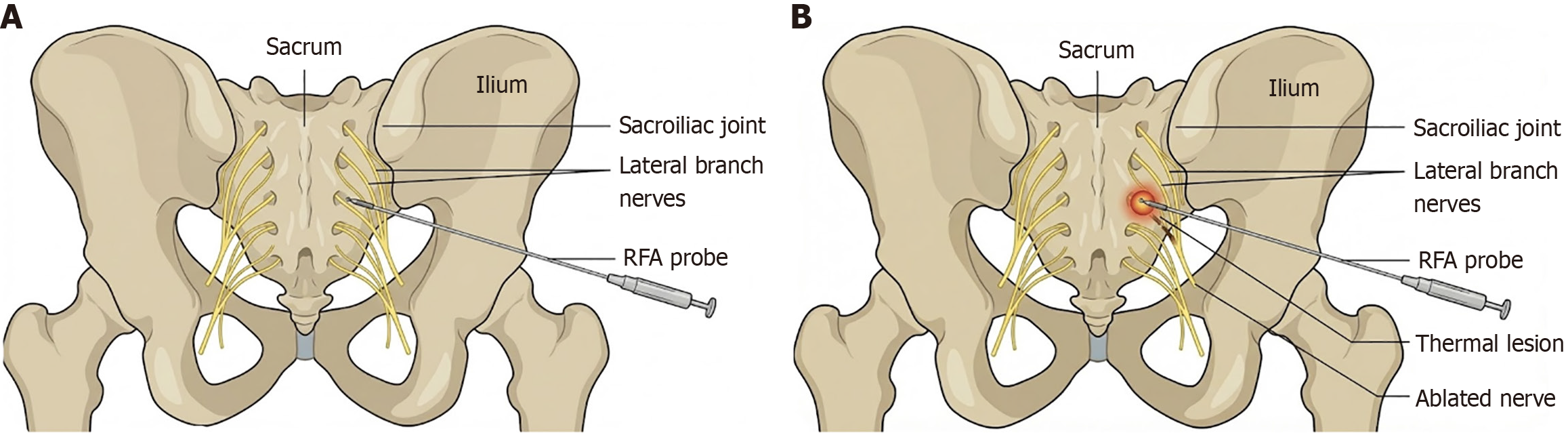
Figure 6 This medical illustration compares the sacroiliac joint pre and post radiofrequency ablation.
A: This image shows the correct posterior anatomical view of the sacrum and ilium. The lateral branches of the posterior sacral nerves are visible, and a radiofrequency ablation probe is positioned on one of them, ready for the procedure; B: This image shows the same posterior view after the radiofrequency ablation procedure has been performed. A thermal lesion is now visible at the tip of the probe, and the targeted nerve is ablated, interrupting the pain signals. RFA: Radiofrequency ablation.
- Citation: Soin A, Mandava A, Soin G, Kloth D, Staats P, Kalia H, Kim P. Sacroiliac joint injections and radiofrequency ablation for pain management: A clinical review. World J Orthop 2026; 17(6): 120581
- URL: https://www.wjgnet.com/2218-5836/full/v17/i6/120581.htm
- DOI: https://dx.doi.org/10.5312/wjo.v17.i6.120581