
















Copyright: ©Author(s) 2026.
World J Orthop. Jun 18, 2026; 17(6): 120581
Published online Jun 18, 2026. doi: 10.5312/wjo.v17.i6.120581
Published online Jun 18, 2026. doi: 10.5312/wjo.v17.i6.120581
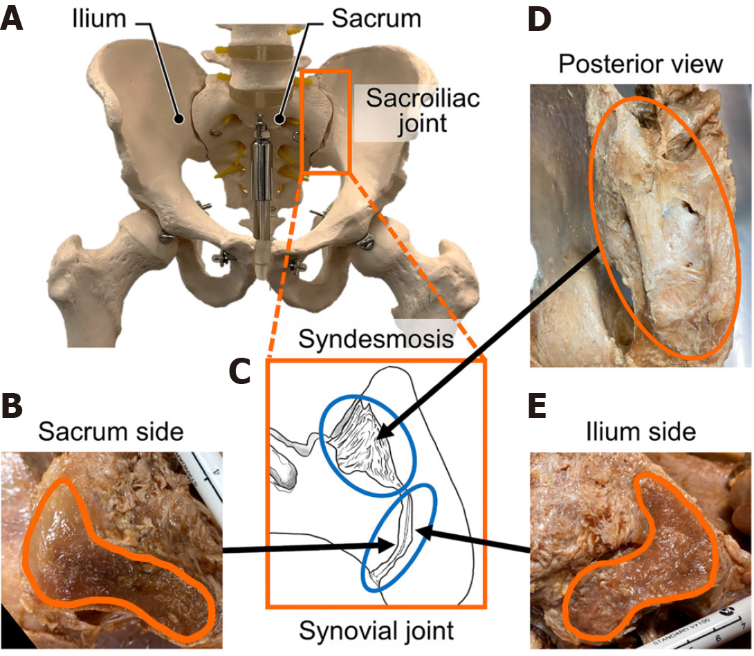
Figure 1 Joint structure[10].
A: The sacroiliac joint is in a position, where joint lines run parallel to the line of gravity; B: The sacral articular surface; C: The cross-sectional image of joint, shows posterior and anterior regions are syndesmoses and synovial joints; D: Syndesmosis, which is composed of sacrospinous ligaments, posterior sacroiliac ligaments, iliolumbar ligaments, and scarotuberous ligaments from posterior side; E: The ilium articular surface. Meanwhile, the redlines on both sacrum and ilium side indicate the synovial regions. Citations: Toyohara R, Ohashi T. A literature review of biomechanical studies on physiological and pathological sacroiliac joints: Articular surface structure, joint motion, dysfunction and treatments. Clin Biomech (Bristol) 2024; 114: 106233. Copyright ©The Author(s) 2024. Published by Elsevier Ltd. The article is open access (Supplementary material).
- Citation: Soin A, Mandava A, Soin G, Kloth D, Staats P, Kalia H, Kim P. Sacroiliac joint injections and radiofrequency ablation for pain management: A clinical review. World J Orthop 2026; 17(6): 120581
- URL: https://www.wjgnet.com/2218-5836/full/v17/i6/120581.htm
- DOI: https://dx.doi.org/10.5312/wjo.v17.i6.120581