
















Copyright: ©Author(s) 2026.
World J Orthop. May 18, 2026; 17(5): 118163
Published online May 18, 2026. doi: 10.5312/wjo.v17.i5.118163
Published online May 18, 2026. doi: 10.5312/wjo.v17.i5.118163
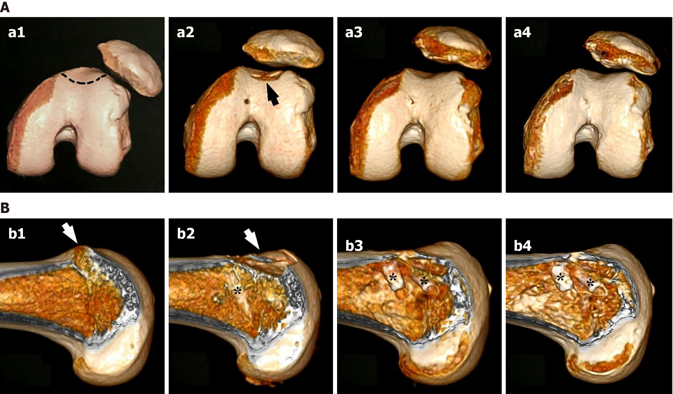
Figure 2 Preoperative and postoperative three-dimensional computed tomography images of a case of left knee recurrent patellar dislocation with type D Dejour trochlear dysplasia.
A: Before surgery, a spur on the proximal side of the trochlear groove and subluxation of the patella could be seen. The black-dashed line was the plan for the new groove (a1); two days after precise arthroscopic mini-trochleoplasty (PAM trochleoplasty), the spur was removed, and two pieces of subchondral bone attached to the osteochondral flap could be seen on the new groove (black arrow). Reduction of the patella to a normal position fit for a new groove showed good patellofemoral congruence (a2); subchondral bone healing resulted in a smooth groove at 7 months after surgery (a3); bone remodeling of the groove and better patellofemoral congruence at 2 years after surgery (a4); B: Before surgery, a sagittal image illustrated the spur (white arrow) on the proximal side of the groove (b1); two days after PAM trochleoplasty, the spur and part of the trochlear bump were removed (white arrow), and subchondral bone pieces attached to the osteochondral flap could be seen (b2); bone healing of the subchondral bone formed a smooth groove at 7 months after surgery (b3); bone remodeling of the new groove at 2 years after surgery (b4). *: Lateral row anchors of suture bridge for fixation of osteochondral flap.
- Citation: Xu H, Ding M, Liao BH, Lei SG, Zheng C, Wang YC. Precise arthroscopic mini-trochleoplasty combined with medial patellofemoral ligament reconstruction yields comparable complications to isolated medial patellofemoral ligament reconstruction. World J Orthop 2026; 17(5): 118163
- URL: https://www.wjgnet.com/2218-5836/full/v17/i5/118163.htm
- DOI: https://dx.doi.org/10.5312/wjo.v17.i5.118163