
















Copyright: ©Author(s) 2026.
World J Orthop. Apr 18, 2026; 17(4): 117533
Published online Apr 18, 2026. doi: 10.5312/wjo.v17.i4.117533
Published online Apr 18, 2026. doi: 10.5312/wjo.v17.i4.117533
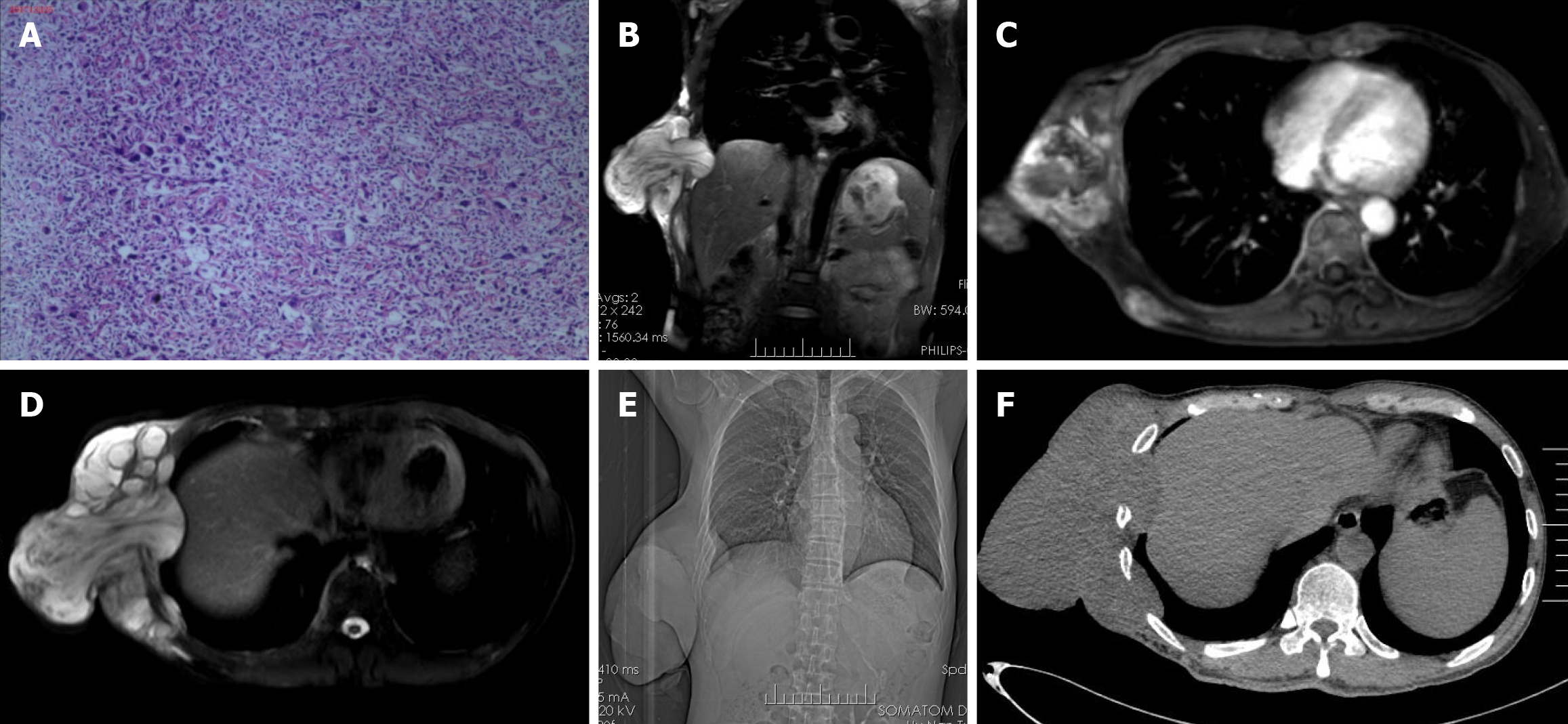
Figure 1 computed tomography, magnetic resonance imaging and histological images of the lesion.
A: Hematoxylin and eosin staining of the lesion (10 ×) representing a large population of heterocyst; B: Coronal magnetic resonance imaging (MRI); C and D: Cross-sectional MRI scan; E: Scout view; F: Cross-sectional computed tomography scan indicate a huge irregular lesion locating at the right lower chest wall, part of the lesion invaded into chest cavity while with intact membrane.
- Citation: Li XA, Chen JY, Bu J, Wu HW. Customized three-dimensional printed titanium ribs combined with bilateral flaps for reconstruction of chest wall defect: A case report. World J Orthop 2026; 17(4): 117533
- URL: https://www.wjgnet.com/2218-5836/full/v17/i4/117533.htm
- DOI: https://dx.doi.org/10.5312/wjo.v17.i4.117533