
















Copyright: ©Author(s) 2026.
World J Orthop. Apr 18, 2026; 17(4): 114757
Published online Apr 18, 2026. doi: 10.5312/wjo.v17.i4.114757
Published online Apr 18, 2026. doi: 10.5312/wjo.v17.i4.114757
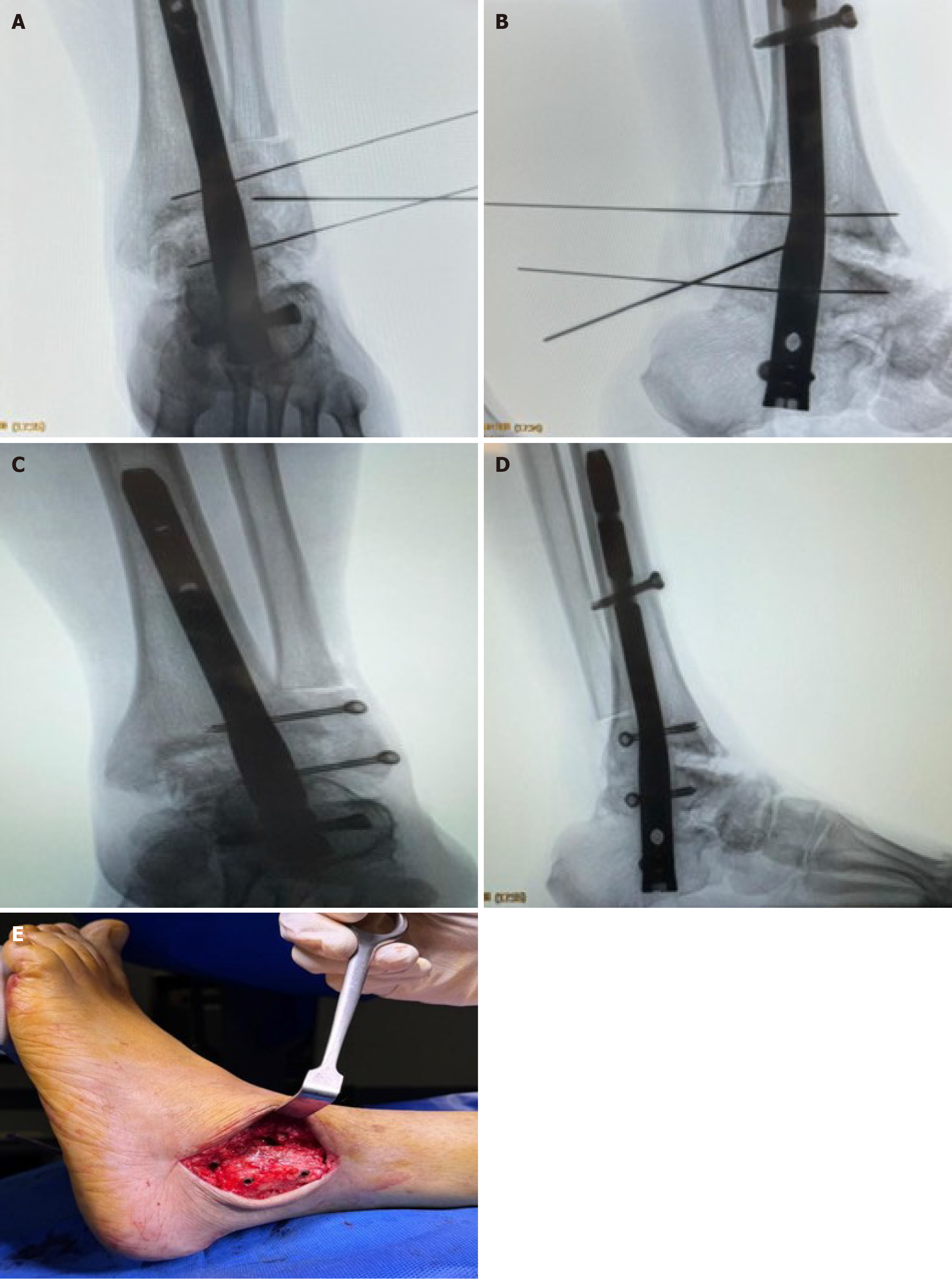
Figure 6 Fibular reduction and fixation.
A: Anteroposterior radiographic view showing fibular reduction with cannulated wires; B: Lateral radiographic view with cannulated wires; C: Anteroposterior radiographic view showing screw insertion; D: Lateral radiographic view showing screw placement; E: Final clinical image showing two screws inserted from fibula creating the “trap door” construct.
- Citation: Elalfy MM, Embaby OM, Abulaban OA, Aladl AN, Abdelrazek M, Youssef K, Abushal MH, Kassab F. Transmalleolar tibiotalocalcaneal fusion with biologic augmentation and fibular preservation for chronic Charcot ankle arthropathy: A retrospective cohort study. World J Orthop 2026; 17(4): 114757
- URL: https://www.wjgnet.com/2218-5836/full/v17/i4/114757.htm
- DOI: https://dx.doi.org/10.5312/wjo.v17.i4.114757