
















Copyright: ©Author(s) 2026.
World J Orthop. Mar 18, 2026; 17(3): 117393
Published online Mar 18, 2026. doi: 10.5312/wjo.v17.i3.117393
Published online Mar 18, 2026. doi: 10.5312/wjo.v17.i3.117393
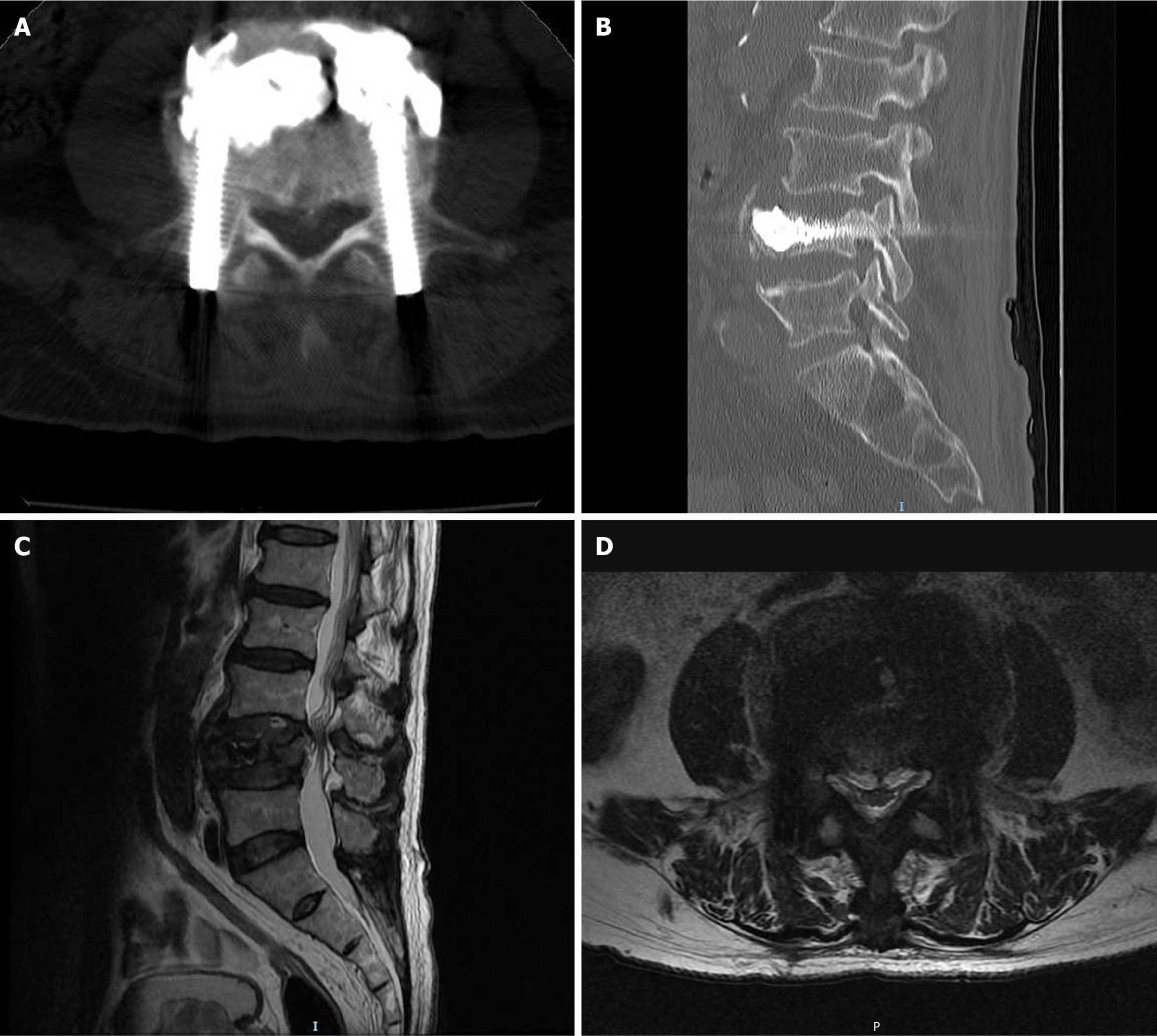
Figure 4 Lumbar computed tomography and magnetic resonance imaging images obtained at the follow-up examination three months after surgery.
A and B: Postoperative three-month lumbar spine axial (A) and sagittal (B) computed tomography images; C: Postoperative three-month sagittal short tau inversion recovery image of lumbar spine magnetic resonance imaging; D: Postoperative three-month lumbar magnetic resonance imaging transverse section image acquired at the lower margin of the L3/4 intervertebral disc level.
- Citation: Long JQ, Yang H, Cheng YW, Shen T, Zou JY, Zhang GW. Percutaneous vertebroplasty and compression screw fixation for osteoporotic vertebral fractures with bilateral pedicle fractures: A case report. World J Orthop 2026; 17(3): 117393
- URL: https://www.wjgnet.com/2218-5836/full/v17/i3/117393.htm
- DOI: https://dx.doi.org/10.5312/wjo.v17.i3.117393