
















Copyright: ©Author(s) 2026.
World J Orthop. Mar 18, 2026; 17(3): 115770
Published online Mar 18, 2026. doi: 10.5312/wjo.v17.i3.115770
Published online Mar 18, 2026. doi: 10.5312/wjo.v17.i3.115770
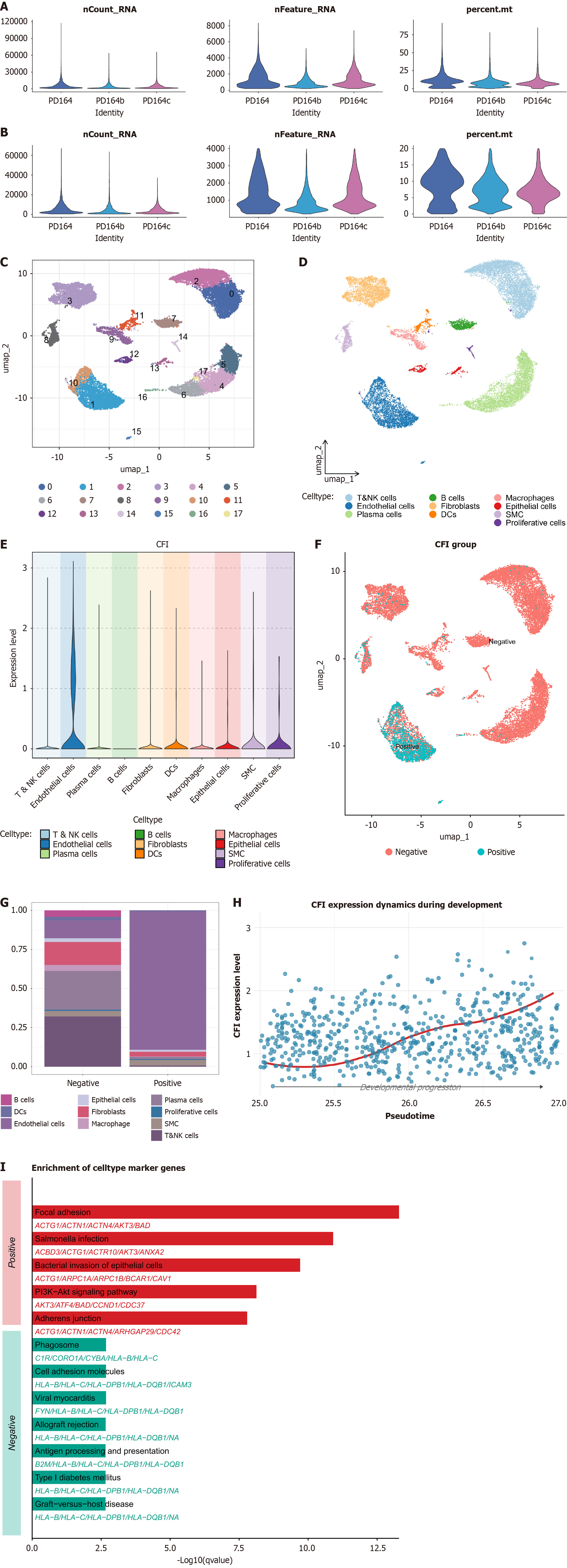
Figure 5 Single-cell characterization of complement factor I expression and functional enrichment in periodontitis.
A and B: Quality control of single cell data; C-E: Expression distribution of complement factor I (CFI) across different cell types; F and G: Identification of the primary cellular source of CFI; F: Proportion of each cell subset in CFI-positive and CFI-negative groups; G: Comparative analysis demonstrating endothelial cells as the dominant source of CFI expression; H: Cell trajectory analysis showing progressive increase of CFI expression during endothelial cell differentiation; I: Kyoto Encyclopedia of Genes and Genomes pathway enrichment analysis of differentially expressed genes in CFI-positive vs CFI-negative groups, highlighting significant enrichment in pathways related to cell adhesion, bacterial infection, and intracellular signaling.
- Citation: Man YN, Zhong LY, Wen YL, He ML. Machine learning identifies complement factor I as a shared mediator of periodontitis and ossification of posterior longitudinal ligament. World J Orthop 2026; 17(3): 115770
- URL: https://www.wjgnet.com/2218-5836/full/v17/i3/115770.htm
- DOI: https://dx.doi.org/10.5312/wjo.v17.i3.115770