
















Copyright: ©Author(s) 2026.
World J Orthop. Mar 18, 2026; 17(3): 113746
Published online Mar 18, 2026. doi: 10.5312/wjo.v17.i3.113746
Published online Mar 18, 2026. doi: 10.5312/wjo.v17.i3.113746
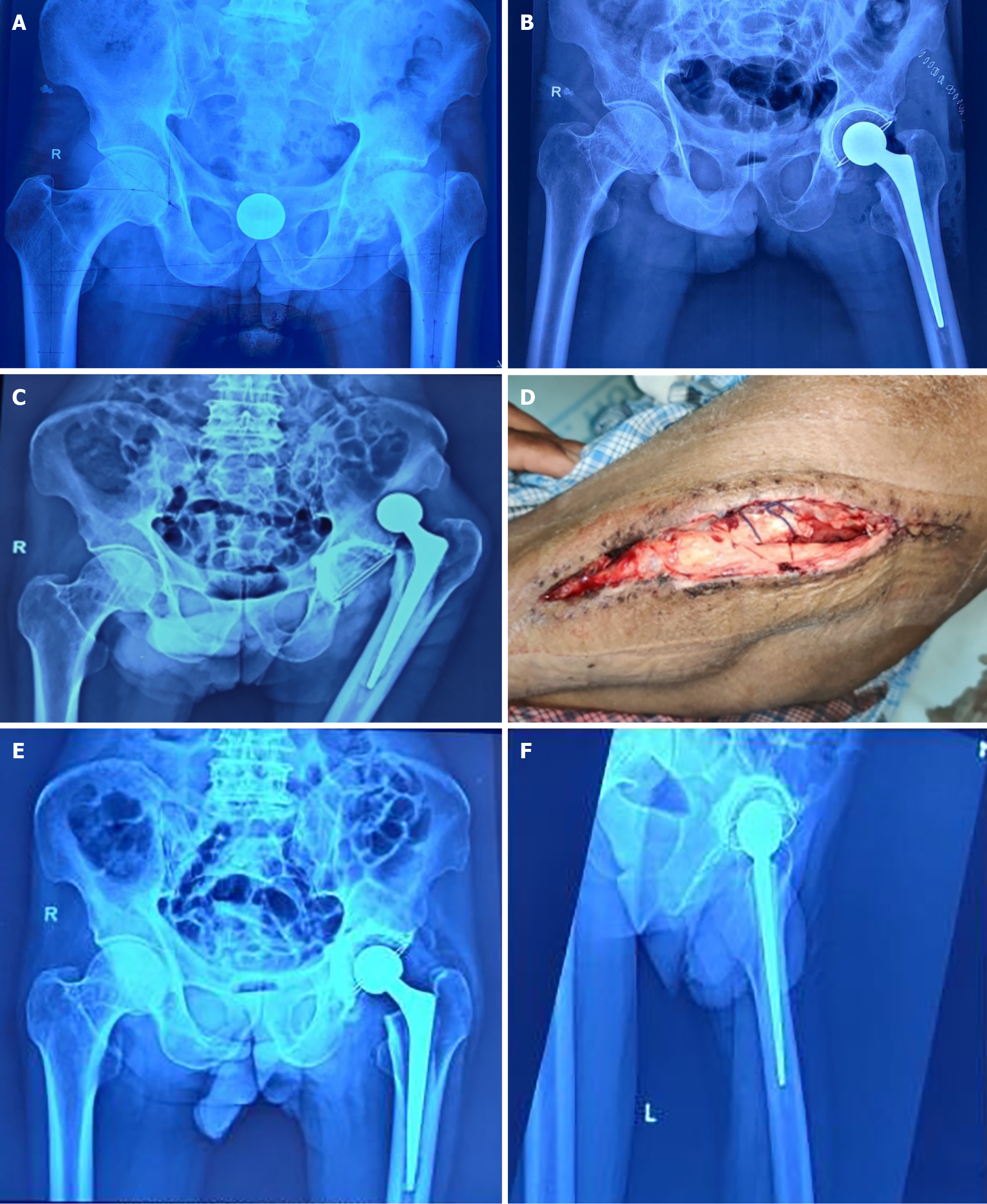
Figure 6 Imaging information of a 55-year-old male with advanced active stage 4 tuberculosis left hip.
A: Pre-operative pelvic radiograph (anteroposterior view) of the patient; B: Immediate post-operative radiograph of the same patient following cemented total hip arthroplasty; C: Patient developed hip dislocation; D: Wound dehiscence in the early post-operative period following a slip on the ground. Closed reduction was done, and the wound was managed conservatively; E and F: Showing satisfactory outcome at 28 months follow-up.
- Citation: Julfiqar M, Bin Sabir A, Bhateja D, Siddiqui YS, Alam A, Shoaib SM, Khan AQ, Asif N, Abbas M, Jilani LZ, Faizan M. Outcome of single-stage total hip arthroplasty in advanced active tubercular arthritis of the hip in Indian patients. World J Orthop 2026; 17(3): 113746
- URL: https://www.wjgnet.com/2218-5836/full/v17/i3/113746.htm
- DOI: https://dx.doi.org/10.5312/wjo.v17.i3.113746