
















Copyright: ©Author(s) 2026.
World J Orthop. Mar 18, 2026; 17(3): 113562
Published online Mar 18, 2026. doi: 10.5312/wjo.v17.i3.113562
Published online Mar 18, 2026. doi: 10.5312/wjo.v17.i3.113562
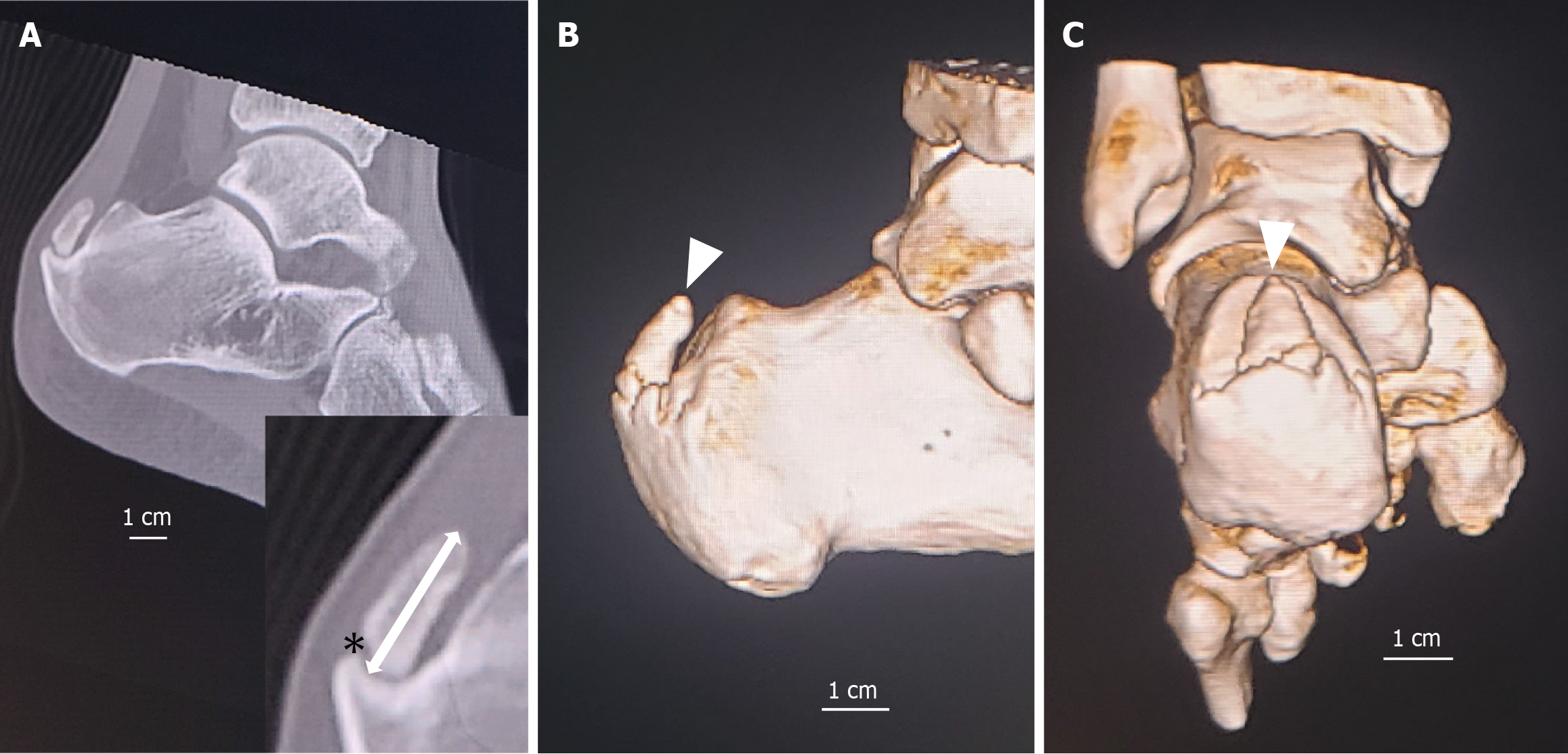
Figure 1 Spur fracture in insertional Achilles tendinopathy.
A: Sagittal computed tomography (CT) image showing a spur fracture (asterisk). Spur length was calculated by subtracting the fracture gap (asterisk) from the total distance between the base and the tip of the spur (arrow); B: Three-dimensional lateral CT image showing spur morphology (arrowhead); C: Three-dimensional posteroanterior CT image showing spur morphology (arrowhead).
- Citation: Nakajima K. Characterizing posterior calcaneal spur fractures in insertional Achilles tendinopathy: A case control study. World J Orthop 2026; 17(3): 113562
- URL: https://www.wjgnet.com/2218-5836/full/v17/i3/113562.htm
- DOI: https://dx.doi.org/10.5312/wjo.v17.i3.113562