
















©The Author(s) 2026.
World J Orthop. Feb 18, 2026; 17(2): 113932
Published online Feb 18, 2026. doi: 10.5312/wjo.v17.i2.113932
Published online Feb 18, 2026. doi: 10.5312/wjo.v17.i2.113932
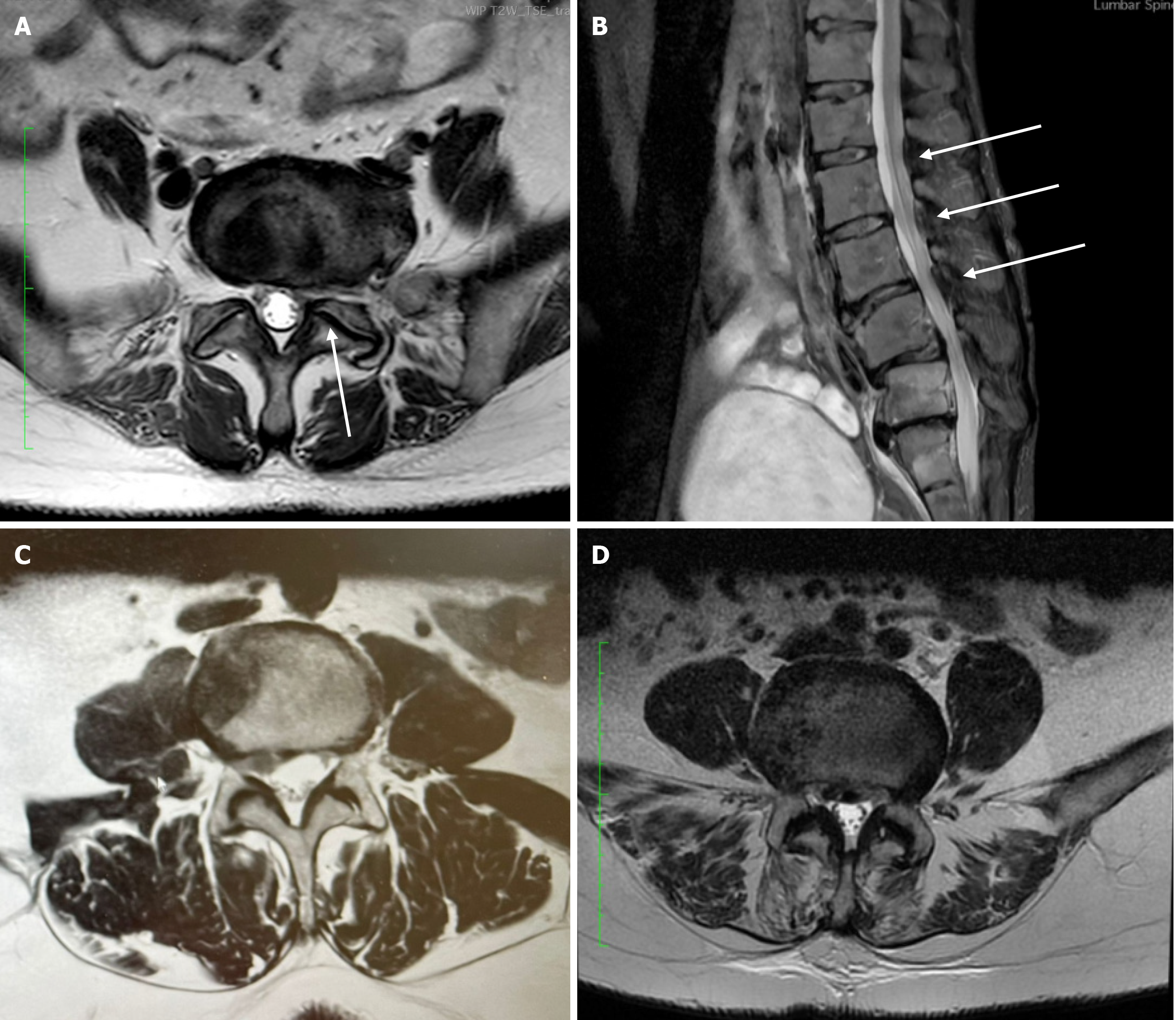
Figure 2 Imaging examples of degenerative changes contributing to central canal stenosis.
A: Axial T2-weighted magnetic resonance imaging (MRI) from a 66-year-old male with unilateral facet joint hypertrophy (white arrow) narrowing the central canal; B: Sagittal T2-weighted MRI from a 49-year-old male showing ligamentum flavum thickening and buckling into the spinal canal (white arrows); C: Axial T2-weighted MRI from a 75-year-old male with lateral recess stenosis; D: Axial T2-weighted MRI from an 82-year-old male with central canal stenosis due to a combination of bilateral facet joint hypertrophy, ligamentum flavum thickening, and a central disc herniation.
- Citation: Chatzivasiliadis M, Konstantinou P, Koulalis D, Kostretzis L, Gkantsinikoudis N, Chaniotakis C, Gkoumousian K, Kapetanakis S. Laminectomy alone vs laminectomy with posterior fusion in lumbar spinal stenosis: The role of instability. World J Orthop 2026; 17(2): 113932
- URL: https://www.wjgnet.com/2218-5836/full/v17/i2/113932.htm
- DOI: https://dx.doi.org/10.5312/wjo.v17.i2.113932