
















©The Author(s) 2026.
World J Orthop. Feb 18, 2026; 17(2): 112889
Published online Feb 18, 2026. doi: 10.5312/wjo.v17.i2.112889
Published online Feb 18, 2026. doi: 10.5312/wjo.v17.i2.112889
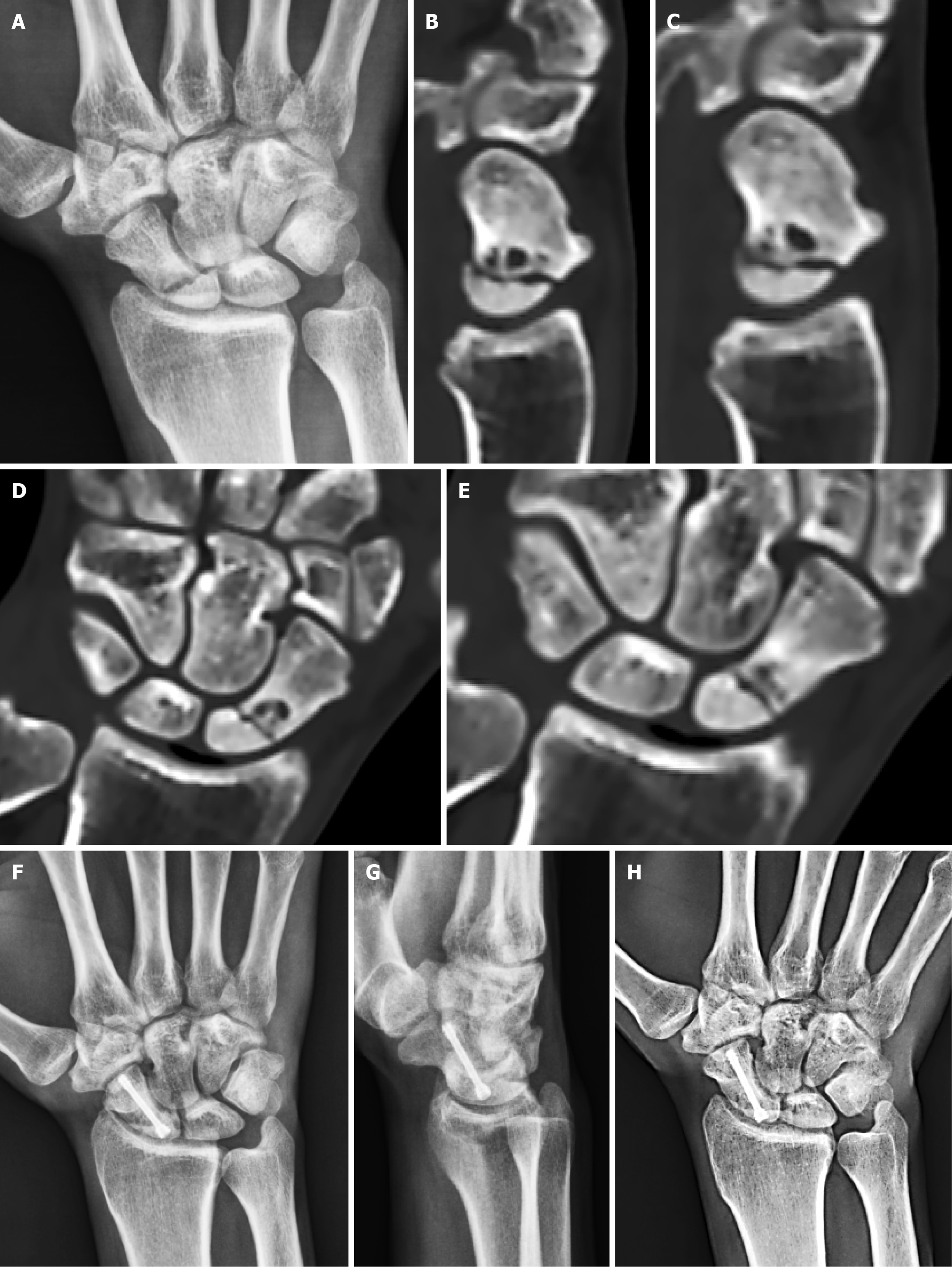
Figure 3 Radiological evaluation of graft-less fixation for proximal pole nonunion.
A: Preoperative anteroposterior wrist radiograph indicating a proximal pole nonunion; B-E: Sagittal and coronal computed tomography slices illustrating the nonunion with cyst changes and bone resorption; F and G: Follow-up radiographs at three-months (anteroposterior and lateral views) demonstrating healing after graft-less fixation; H: Anteroposterior wrist radiograph at five-months confirming full consolidation.
- Citation: Elshahhat A, Ahmed M, Saleh A, Abed Y, Elashmawy H, Nour K. Necessity of grafting in scaphoid nonunion fixation: A comparative outcome analysis. World J Orthop 2026; 17(2): 112889
- URL: https://www.wjgnet.com/2218-5836/full/v17/i2/112889.htm
- DOI: https://dx.doi.org/10.5312/wjo.v17.i2.112889