
















Copyright: ©Author(s) 2026.
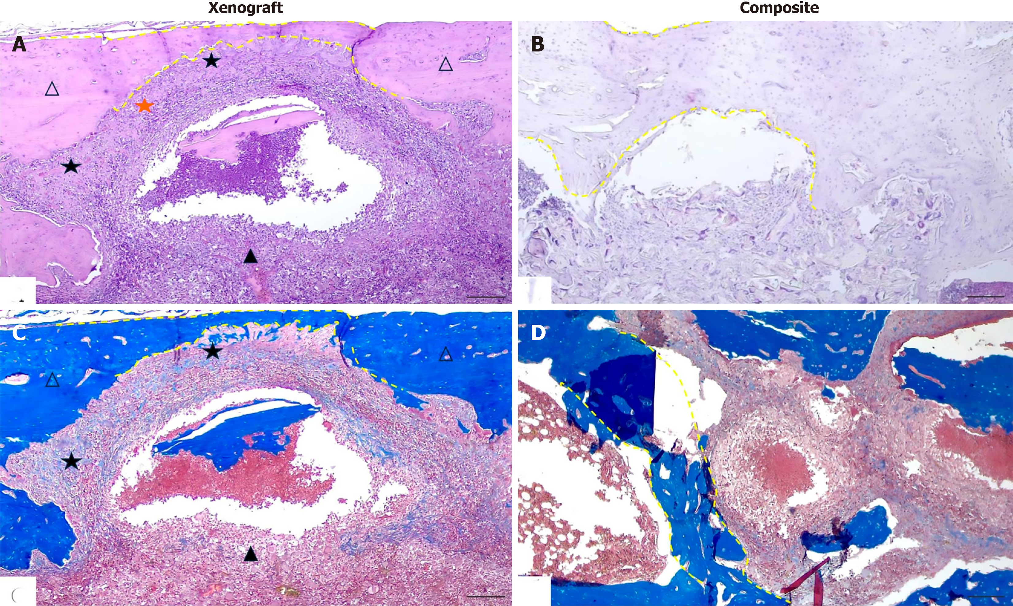
Figure 3 Representative photomicrographs of the cortical plate defect area and the intramedullary space on day 60 after filling the bone defect with xenograft and composite.
A: Xenograft. The defect zone is completely closed by thin newly formed bone tissue (dashed line), consisting of bone trabeculae with prominent lacunae containing osteocytes. Within the intramedullary space, bone xenograft fragments with signs of lysis are identified; B: Composite. The cortical plate defect is fully restored. The composite is undergoing active remodeling and is being replaced by bone tissue; C: Xenograft. The newly formed bone tissue is actively mineralizing. A fragment of remodeling xenograft is present in the intramedullary space; D: Composite. The cortical plate is completely restored. The composite is partially resorbed and is being replaced by mature bone trabeculae. Hematoxylin and eosin staining (A and B). Masson’s trichrome (C and D). Original magnification × 40, scale bar 500 μm. White triangle: Bone plate defect margin. Black triangle: Multinucleated giant cells. Orange five-pointed star: Polymorphonu
- Citation: Tuleubayev BE, Makhatov BK, Vinokurov VA, Kamyshanskiy YK, Kossilova YY, Akhmetova SB, Koshanova AA, Davletbaev MZ, Arutyunyan MG. Osteoreparative potential of nanofibrillated cellulose, xenograft, and butvar-phenolic adhesive-based composite. World J Orthop 2026; 17(11): 118182
- URL: https://www.wjgnet.com/2218-5836/full/v17/i11/118182.htm
- DOI: https://dx.doi.org/10.5312/wjo.118182