
















Copyright: ©Author(s) 2026.
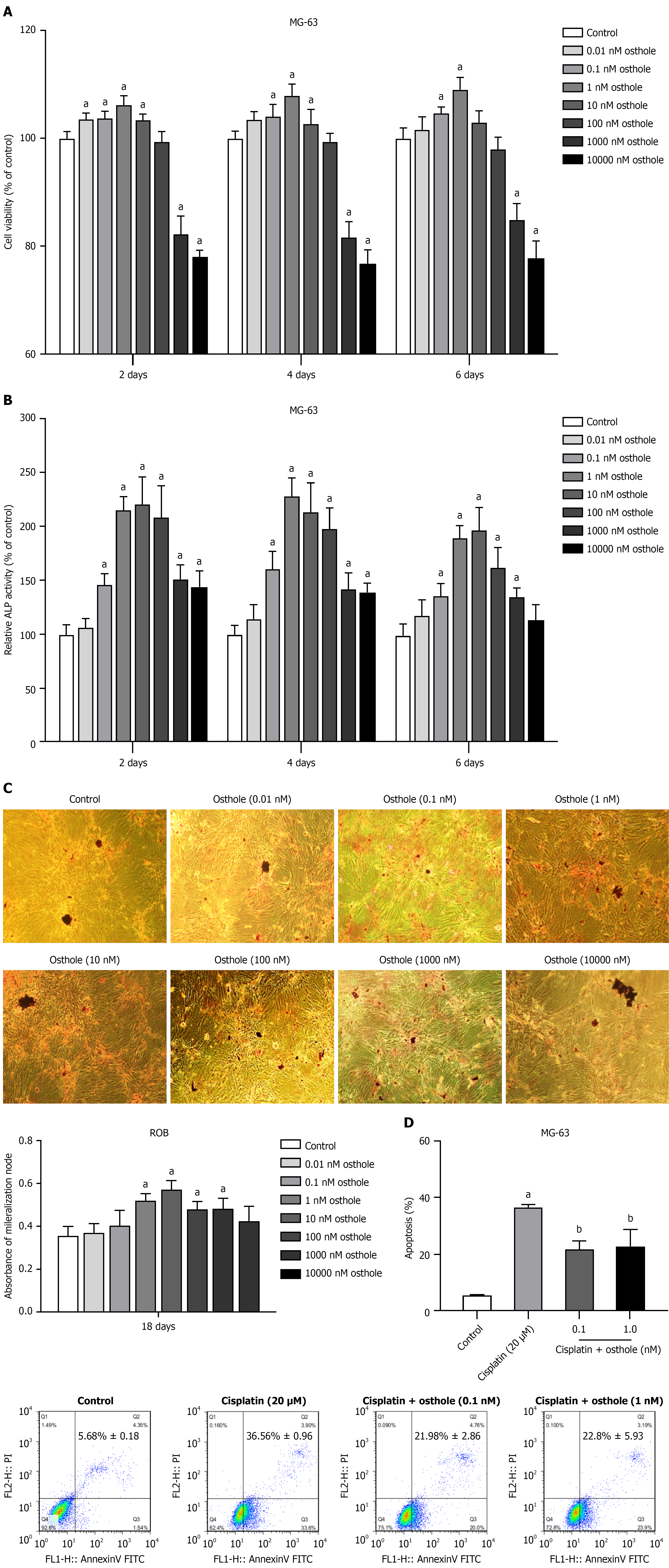
Figure 2 Osthole modulates osteoblastic proliferation, differentiation, mineralization, and apoptosis.
aP < 0.05 vs control (one-way analysis of variance followed by Dunnett’s post-hoc test); bP < 0.05 vs cisplatin alone (one-way analysis of variance with Tukey’s post-hoc test). A: MG-63 cells were treated with different concentrations of osthole (0.01-10000 nM) or vehicle dimethyl sulfoxide in medium containing 1% fetal bovine serum for 2 days, 4 days, and 6 days, and cell proliferation was detected by 3-(4,5-dimethylthiazol-2-yl)-2,5-diphenyltetrazolium bromide assay. Data are mean ± SD (n = 5 independent experiments); B: Alkaline phosphatase(ALP) activity of MG-63 cells treated with the above concentrations of osthole was measured by ALP activity assay. Cell lysates were collected after treatment, and ALP activity was normalized to the absorbance value of 3-(4,5-dimethylthiazol-2-yl)-2,5-diphenyltetrazolium bromide at 490 nm. Data are mean ± SD (n = 5); C: After rat calvarial osteoblasts were cultured with different concentrations (0.01-10000 nM) of osthole for 18 days, the number of mineralized bone nodules was quantified by Alizarin Red S staining (original magnification × 100). The absorbance of the released Alizarin Red S stain was measured at 570 nm. Data are mean ± SD (n = 3); D: The mean percentage of apoptotic MG-63 cells was measured by flow cytometry. Data are mean ± SD (n = 3). ALP: Alkaline phosphatase; ROBs: Rat calvarial osteoblasts; FL1-H: FL1-height; FITC: Fluorescein isothiocyanate.
- Citation: Zhu TY, Yuan M, Wen XC, Tian CC, Han YH, Zheng C, Wu YT, Xia Q, Wang Y. Osthole exerts osteoprotective effect by activating ER, PI3K/AKT, MEK/ERK, and Wnt/β-catenin signaling pathways. World J Orthop 2026; 17(10): 121526
- URL: https://www.wjgnet.com/2218-5836/full/v17/i10/121526.htm
- DOI: https://dx.doi.org/10.5312/wjo.121526