
















©The Author(s) 2026.
World J Orthop. Jan 18, 2026; 17(1): 112262
Published online Jan 18, 2026. doi: 10.5312/wjo.v17.i1.112262
Published online Jan 18, 2026. doi: 10.5312/wjo.v17.i1.112262
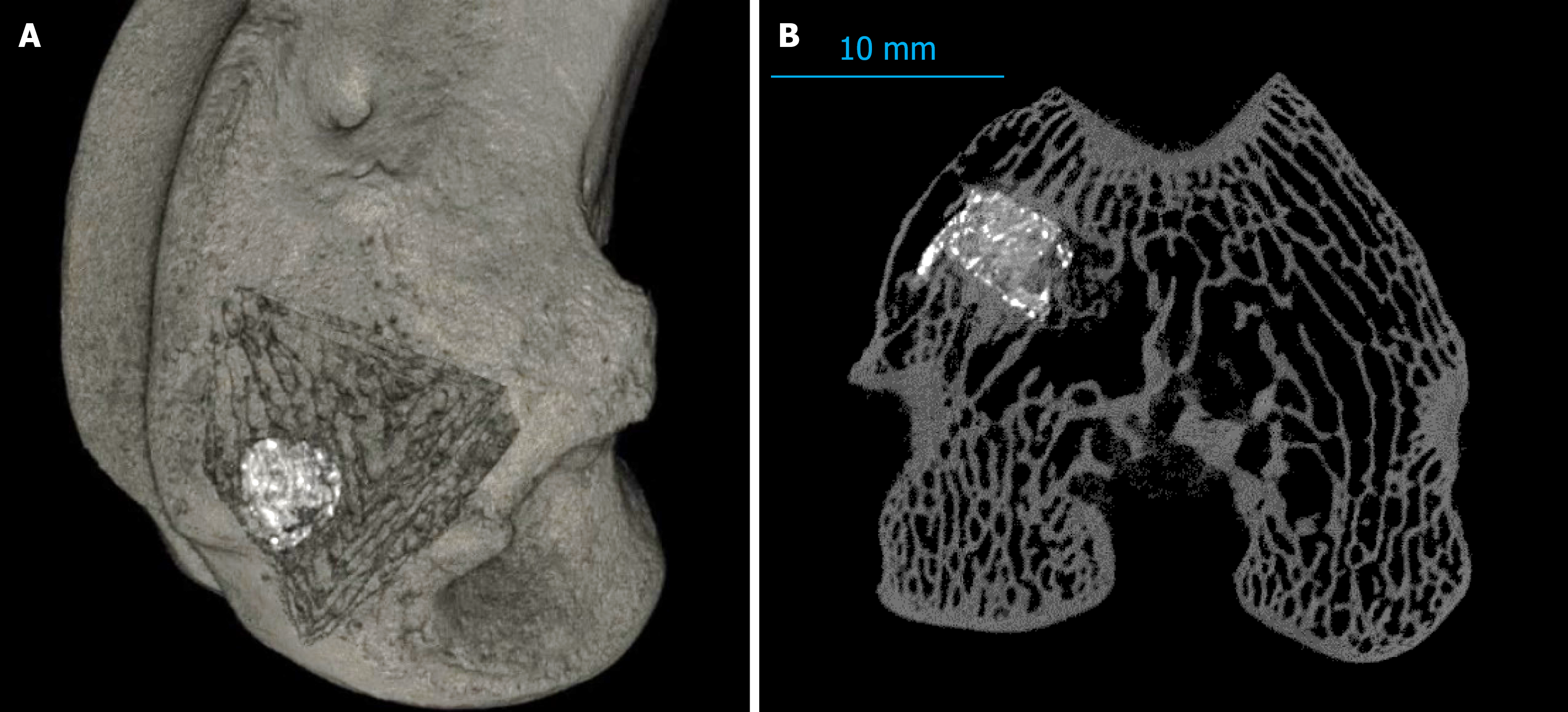
Figure 2 Computed tomography images of the distal part of the right femur of a rabbit from group 4 (autopsy at 6 months control point).
A: Three-dimensional reconstruction showing the heterogeneous density of the implanted material with visible inclusions of calcium sulfate and bone trabeculae ingrowth; B: Computed tomography cross-sectional slice confirming the implant structure and absence of peri-implant resorption.
- Citation: Petukhova VV, Mushkin AY, Maletin AS, Dogonadze MZ, Zabolotnykh NV, Dyakova ME, Esmedlyaeva DS, Vinogradova TI, Kostik MM. Novel use of bisphosphonates to improve surgical outcomes in experimental bone tuberculosis. World J Orthop 2026; 17(1): 112262
- URL: https://www.wjgnet.com/2218-5836/full/v17/i1/112262.htm
- DOI: https://dx.doi.org/10.5312/wjo.v17.i1.112262