
















©The Author(s) 2026.
World J Orthop. Jan 18, 2026; 17(1): 112222
Published online Jan 18, 2026. doi: 10.5312/wjo.v17.i1.112222
Published online Jan 18, 2026. doi: 10.5312/wjo.v17.i1.112222
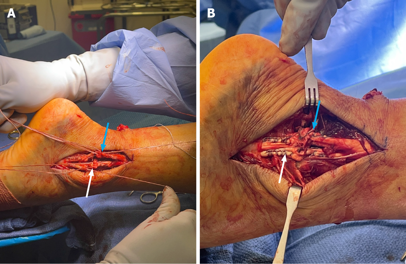
Figure 5 Intraoperative images showing repair of the peroneus longus and brevis tendons.
A: Lacerated peroneus longus (blue arrow) and brevis (white arrow) tendons secured proximally and distally with 2-suture core Kraków repair and manually reapproximated prior to repair; B: Repaired peroneus longus (blue arrow) and brevis (white arrow) tendons visualized from posterior lateral incision.
- Citation: Vlasak A, Orear C, Sakryd G, Metzl J. Achilles, peroneus longus and brevis ruptures with lateral malleolus fracture: A case report and review of literature. World J Orthop 2026; 17(1): 112222
- URL: https://www.wjgnet.com/2218-5836/full/v17/i1/112222.htm
- DOI: https://dx.doi.org/10.5312/wjo.v17.i1.112222