
















©The Author(s) 2026.
World J Orthop. Jan 18, 2026; 17(1): 112222
Published online Jan 18, 2026. doi: 10.5312/wjo.v17.i1.112222
Published online Jan 18, 2026. doi: 10.5312/wjo.v17.i1.112222
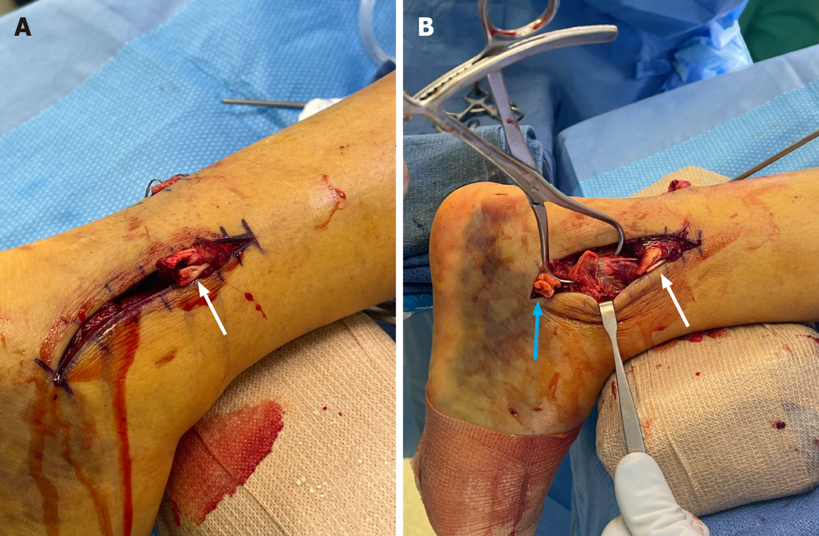
Figure 3 Intraoperative images showing the exposed peroneus tendon lacerations.
A: Posterior-lateral approach over the distal fibula demonstrates lacerations of both the peroneus longus and brevis tendons with proximal tendon stumps exposed. White arrow denotes peroneal tendon stumps; B: Posterior-lateral approach over the distal fibula demonstrates fracture reduced with Weber clamp. Both proximal and distal peroneal tendon stumps can be visualized. White arrow denotes proximal tendon stumps and blue arrow denotes distal stumps.
- Citation: Vlasak A, Orear C, Sakryd G, Metzl J. Achilles, peroneus longus and brevis ruptures with lateral malleolus fracture: A case report and review of literature. World J Orthop 2026; 17(1): 112222
- URL: https://www.wjgnet.com/2218-5836/full/v17/i1/112222.htm
- DOI: https://dx.doi.org/10.5312/wjo.v17.i1.112222