
















©The Author(s) 2026.
World J Orthop. Jan 18, 2026; 17(1): 112222
Published online Jan 18, 2026. doi: 10.5312/wjo.v17.i1.112222
Published online Jan 18, 2026. doi: 10.5312/wjo.v17.i1.112222
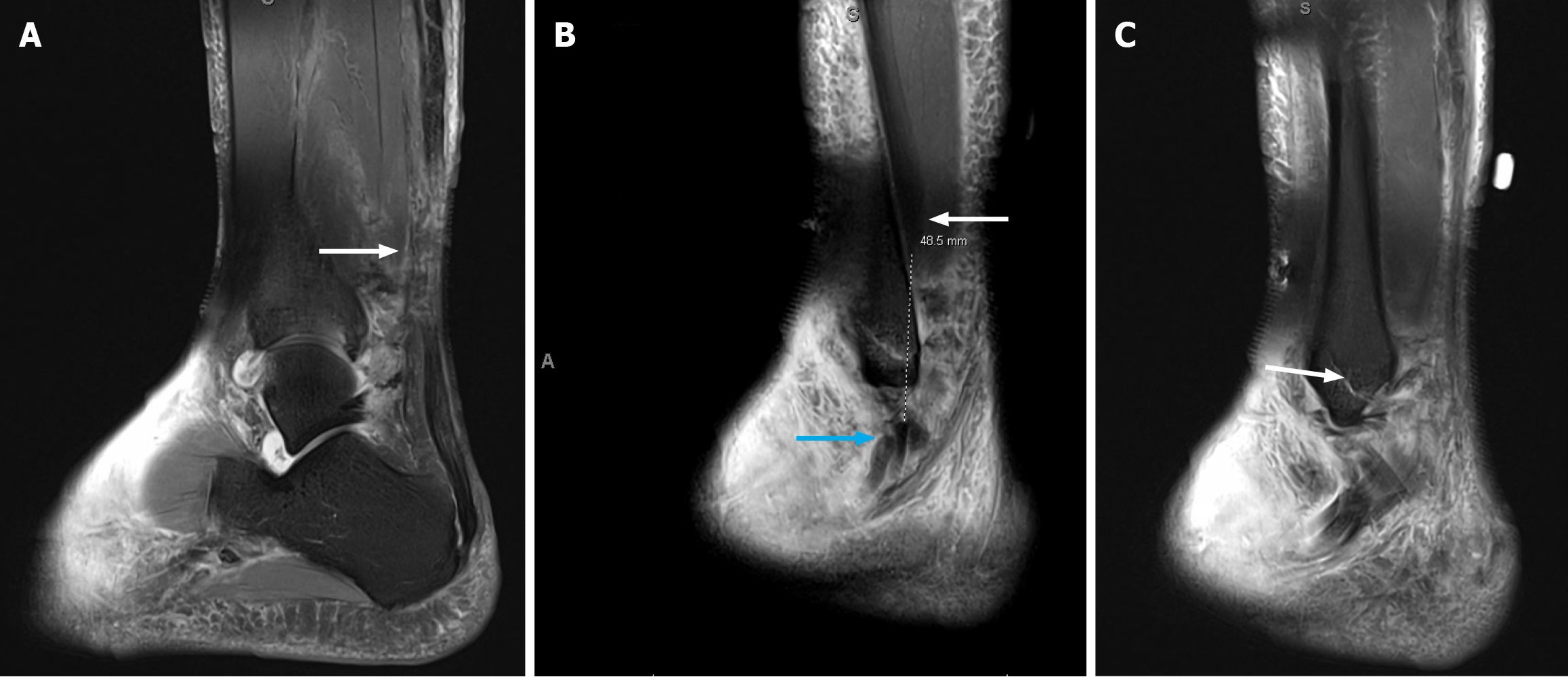
Figure 1 Sagittal spin-echo short inversion time inversion-recovery sequence magnetic resonance imaging of the ankle.
A: Image demonstrating mid substance Achilles rupture and adjacent soft tissue edema. White arrow indicates location of Achilles rupture; B: Image showing complete tears of the peroneus longus and brevis tendons, located just proximal to the lateral malleolus with approximately 5 cm of diastases between the tendon ends. Blue arrow demonstrates distal tendon stumps and white arrow demonstrates proximal tendon substance; C: Image of nondisplaced oblique distal fracture of the fibula. White arrow denotes the fracture.
- Citation: Vlasak A, Orear C, Sakryd G, Metzl J. Achilles, peroneus longus and brevis ruptures with lateral malleolus fracture: A case report and review of literature. World J Orthop 2026; 17(1): 112222
- URL: https://www.wjgnet.com/2218-5836/full/v17/i1/112222.htm
- DOI: https://dx.doi.org/10.5312/wjo.v17.i1.112222