Copyright
©The Author(s) 2025.
World J Orthop. Aug 18, 2025; 16(8): 107083
Published online Aug 18, 2025. doi: 10.5312/wjo.v16.i8.107083
Published online Aug 18, 2025. doi: 10.5312/wjo.v16.i8.107083
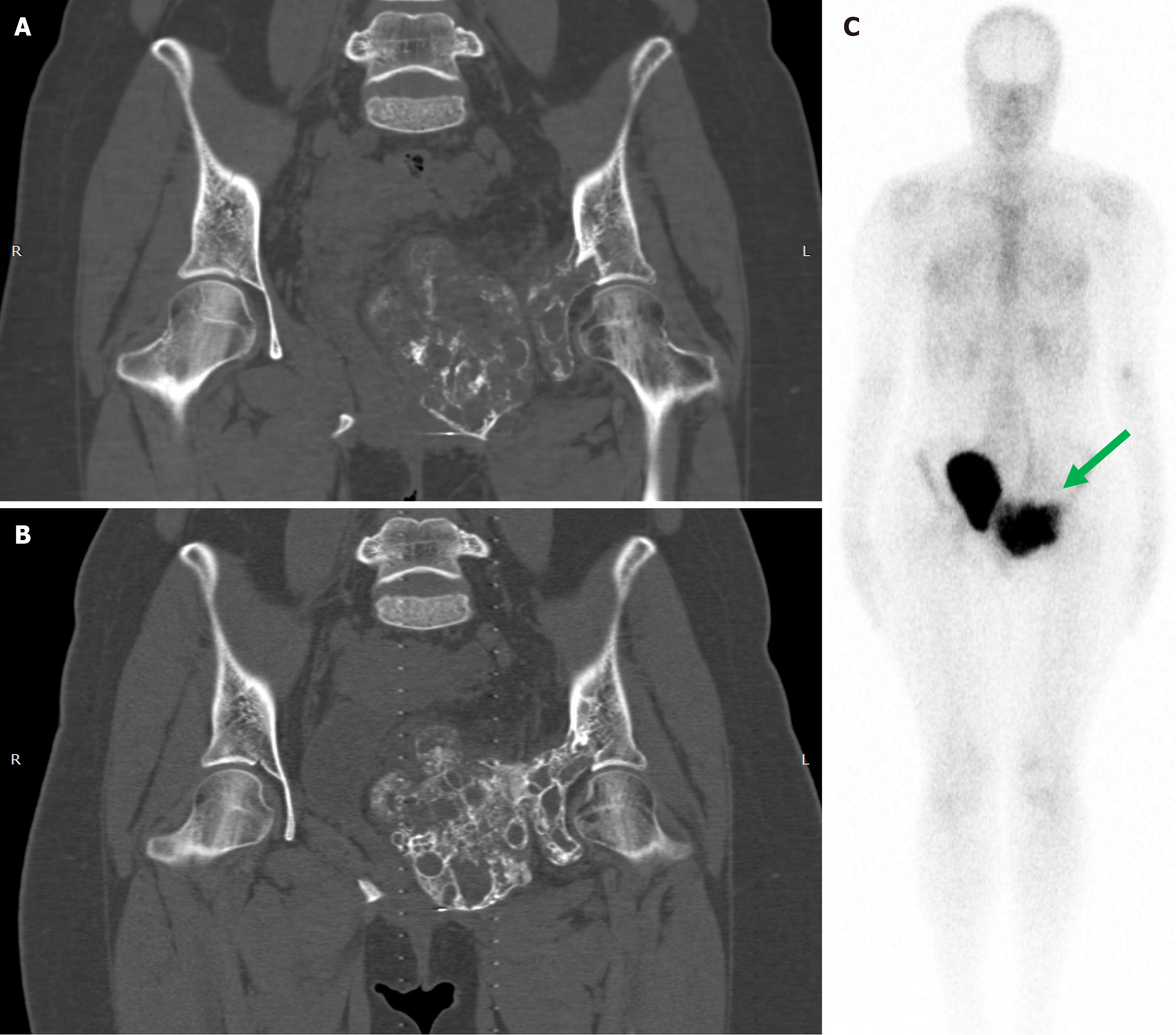
Figure 2 Aneurysmal bone cysts of the left pelvic bones.
A: Frontal computed tomography (CT) scans before denosumab therapy (DT); B: Frontal CT scans 12 months after initiation of DT demonstrate more pronounced ossification and demarcation of the tumor margin; C: Planar bone scintigraphy after DT showing intensive tracer uptake in the ossified tumor (arrow). Bladder accumulating excreted 99mTc-BP is dislocated to the right.
- Citation: Machak GN, Bruland ØS, Kovalev AV, Rodionova SS. Rethinking the role of bisphosphonates after denosumab treatment in locally advanced or unresectable aneurysmal bone cysts: A meta-analysis. World J Orthop 2025; 16(8): 107083
- URL: https://www.wjgnet.com/2218-5836/full/v16/i8/107083.htm
- DOI: https://dx.doi.org/10.5312/wjo.v16.i8.107083


