Copyright
©The Author(s) 2025.
World J Orthop. Aug 18, 2025; 16(8): 106982
Published online Aug 18, 2025. doi: 10.5312/wjo.v16.i8.106982
Published online Aug 18, 2025. doi: 10.5312/wjo.v16.i8.106982
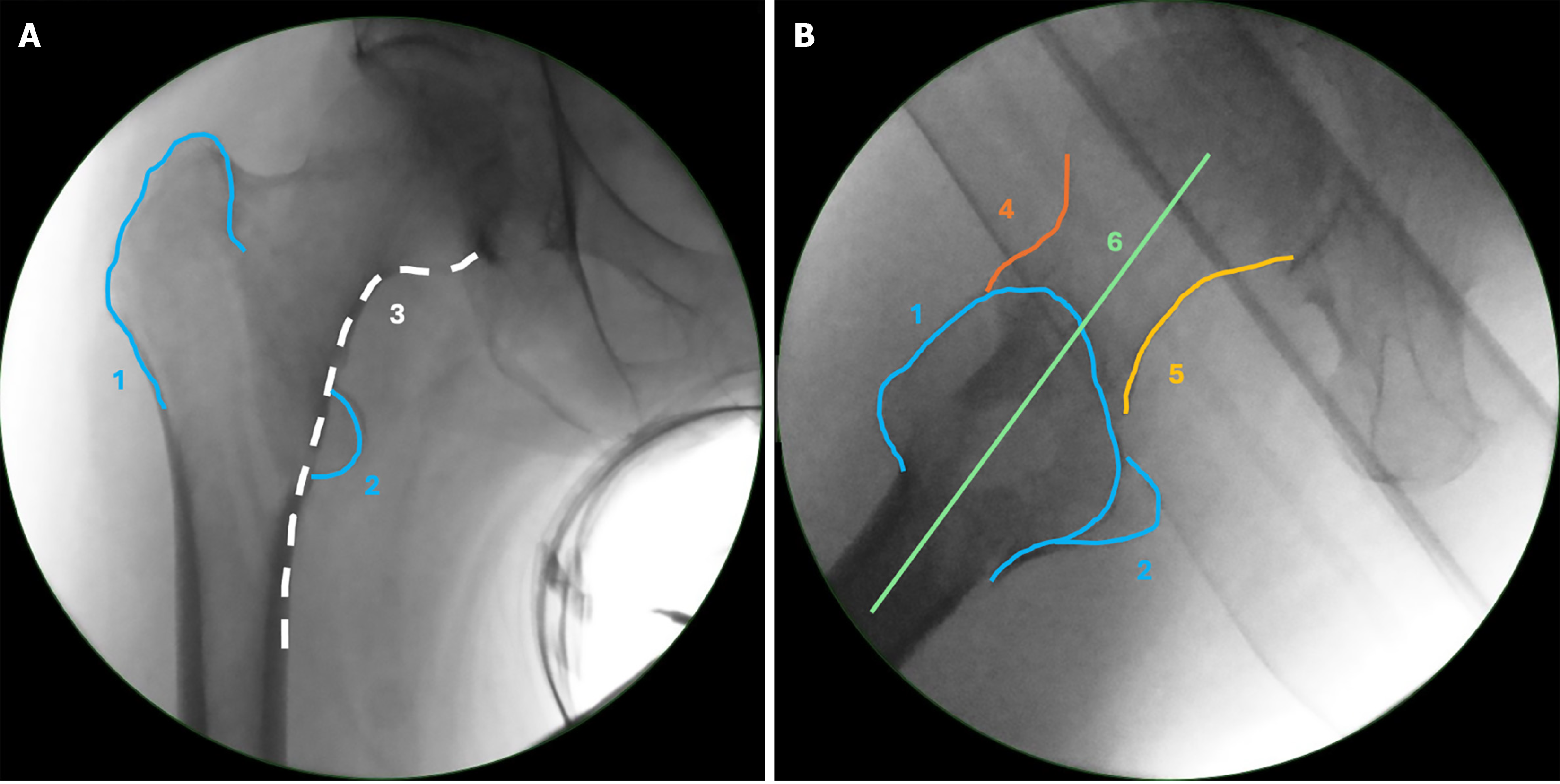
Figure 1 Anatomical landmarks and lines of the proximal femur in anteroposterior and lateral view.
A: Anteroposterior view; B: Lateral view. Greater trochanter (1), lesser trochanter (2), Calcar line (3), anterior line (4), posterior line (5), head-neck-shaft line (6).
- Citation: Wittauer M, Henry J, Sánchez-Rosenberg G, Lambers AP, Jones CW, Yates PJ. Evaluation of reduction quality and implant positioning in intertrochanteric fracture fixation: A review of key radiographic parameters. World J Orthop 2025; 16(8): 106982
- URL: https://www.wjgnet.com/2218-5836/full/v16/i8/106982.htm
- DOI: https://dx.doi.org/10.5312/wjo.v16.i8.106982


