Copyright
©The Author(s) 2025.
World J Orthop. Jul 18, 2025; 16(7): 107913
Published online Jul 18, 2025. doi: 10.5312/wjo.v16.i7.107913
Published online Jul 18, 2025. doi: 10.5312/wjo.v16.i7.107913
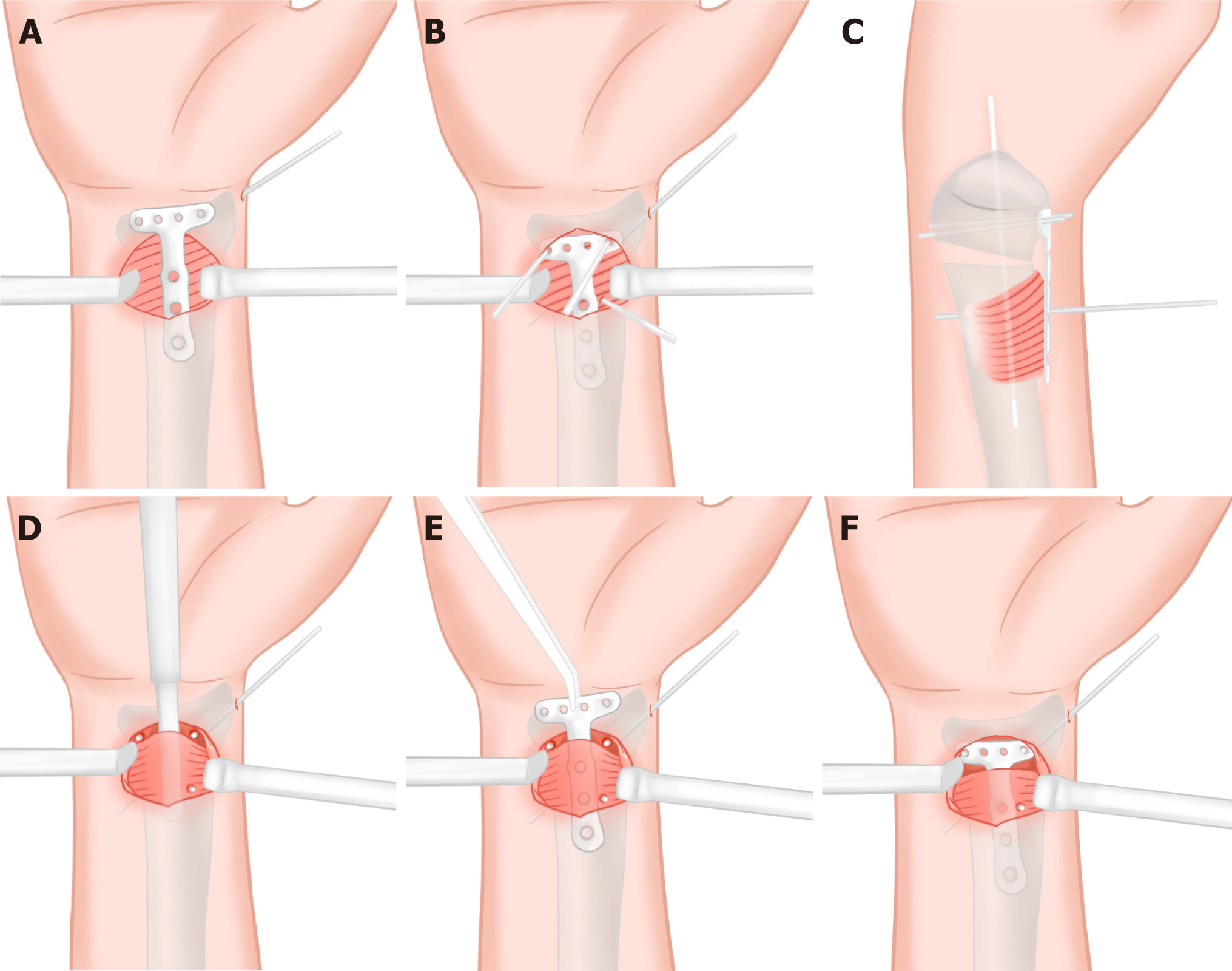
Figure 3 Schematic diagram of the three-point positioning technique.
A: The plate was placed above the unseparated pronator quadratus (PQ) muscle; B and C: Two 1.5 mm Kirschner wires were inserted through the radial and ulnar distal screw holes of the plate, penetrating both cortices. An additional 1.5 mm Kirschner wire was placed adjacent to the ulnar border of the plate's central portion to verify proper radial axis alignment; D: Following trimming of all three Kirschner wires to approximately 0.5 cm in length and leaving them in situ, the plate was removed and subperiosteal dissection of the PQ muscle was performed; E and F: The locking hole of the plate's distal radial and ulnar sides were "guided onto" into the two shorted Kirschner wire posts at the distal end after the plate was inserted into the tunnel from the radial edge of the proximal Kirschner wire.
- Citation: Ye YY, Shen ZQ, Wu CL, Lin YB. Minimally invasive plate osteosynthesis for distal radius fractures using a 3-point positioning technique. World J Orthop 2025; 16(7): 107913
- URL: https://www.wjgnet.com/2218-5836/full/v16/i7/107913.htm
- DOI: https://dx.doi.org/10.5312/wjo.v16.i7.107913


