Copyright
©The Author(s) 2025.
World J Orthop. Jul 18, 2025; 16(7): 107913
Published online Jul 18, 2025. doi: 10.5312/wjo.v16.i7.107913
Published online Jul 18, 2025. doi: 10.5312/wjo.v16.i7.107913
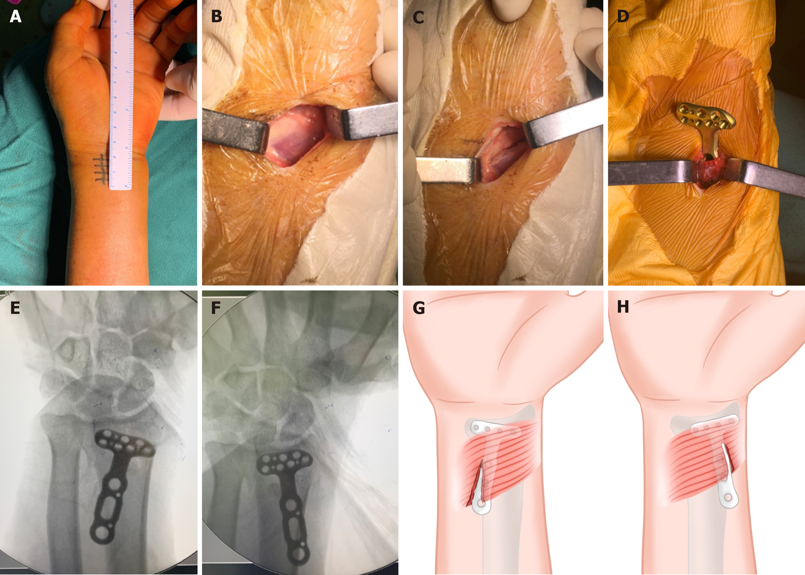
Figure 2 Schematic diagram of injury to the pronator quadratus during minimally invasive plate osteosynthesis with a small incision.
A: The surgical incision; B: The distal margin of the pronator quadratus (PQ) was exposed; C: The PQ was incised; D: The plate was inserted beneath the PQ; E and F: Due to blind penetration, the plate may be placed on the radial or ulnar side during the operation; G and H: The PQ muscle was incised at both its origin and insertion points along the volar surface of the distal third of the radius.
- Citation: Ye YY, Shen ZQ, Wu CL, Lin YB. Minimally invasive plate osteosynthesis for distal radius fractures using a 3-point positioning technique. World J Orthop 2025; 16(7): 107913
- URL: https://www.wjgnet.com/2218-5836/full/v16/i7/107913.htm
- DOI: https://dx.doi.org/10.5312/wjo.v16.i7.107913


