Copyright
©The Author(s) 2025.
World J Orthop. Jul 18, 2025; 16(7): 106570
Published online Jul 18, 2025. doi: 10.5312/wjo.v16.i7.106570
Published online Jul 18, 2025. doi: 10.5312/wjo.v16.i7.106570
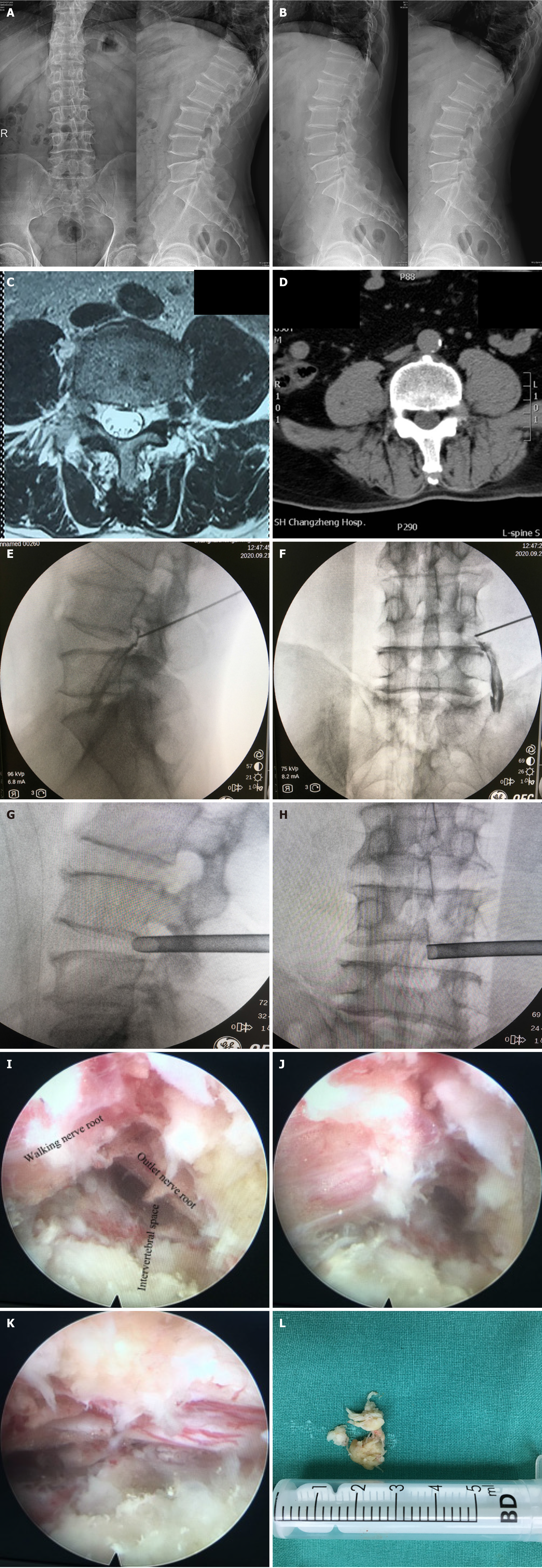
Figure 4 A typical case of selective nerve root block combined with percutaneous transforaminal endoscopic discectomy for treating far lateral lumbar disc herniation.
A56-year-old man was admitted to the hospital with radiating pain in his leftlower limb for 3 months. The diagnosis was far lateral lumbar disc herniation, and the responsible segment and nerve root were the L4/5 and L4 nerve roots. A: The X-ray positive and lateral position of the lumbar spinebefore operation; B: The X-ray hyperextension and flexion position of thelumbar spine before operation; C and D: Magnetic resonance imaging and computed tomography before the operation; E and F: The patients undergoing selective nerve root block before surgery; G and H: X-raypositive and lateral images of channel placement during percutaneous transforaminal endoscopic discectomy operation; I: The microscopic images of the outlet nerve root, walking nerve root and intervertebral space during theoperation; J: The outlet nerve root after decompression; K: The walking nerve heel afterdecompression; L: The nucleus pulposus tissue removed during the operation.
- Citation: Xiao B, Gu X, Zhang JY, Ye XJ, Xi YH, Xu GH, Wang WH. Minimally invasive treatment of far lateral lumbar disc herniation: Selective nerve root block with percutaneous transforaminal endoscopic discectomy. World J Orthop 2025; 16(7): 106570
- URL: https://www.wjgnet.com/2218-5836/full/v16/i7/106570.htm
- DOI: https://dx.doi.org/10.5312/wjo.v16.i7.106570


