Copyright
©The Author(s) 2025.
World J Orthop. Jul 18, 2025; 16(7): 106570
Published online Jul 18, 2025. doi: 10.5312/wjo.v16.i7.106570
Published online Jul 18, 2025. doi: 10.5312/wjo.v16.i7.106570
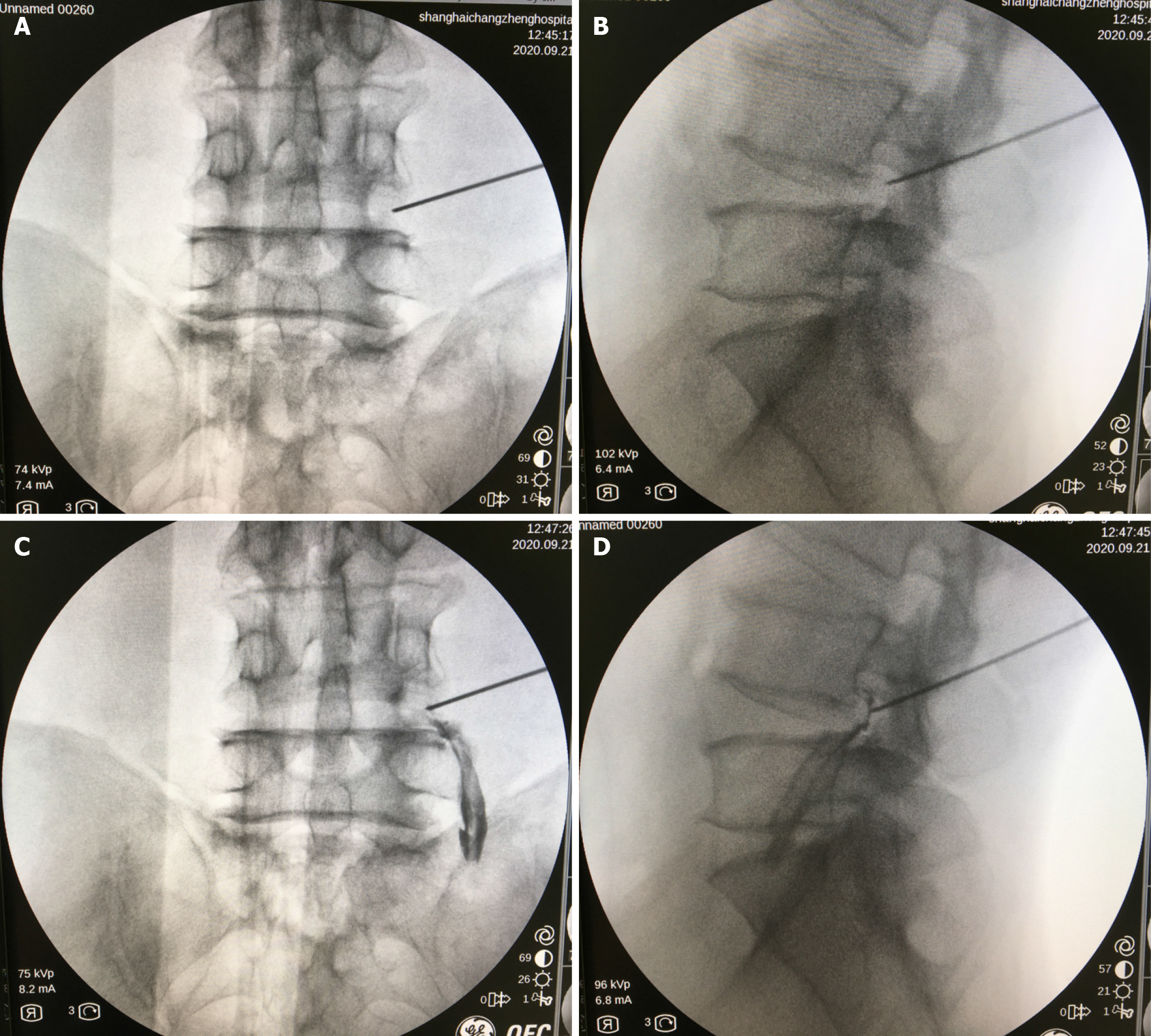
Figure 3 Standardization of selective nerve root block.
A and B: Location of the puncture needle using an X-ray; C and D: Nerve root imaging after the injection of 0.5 mL of iohexol contrast agent around the root.
- Citation: Xiao B, Gu X, Zhang JY, Ye XJ, Xi YH, Xu GH, Wang WH. Minimally invasive treatment of far lateral lumbar disc herniation: Selective nerve root block with percutaneous transforaminal endoscopic discectomy. World J Orthop 2025; 16(7): 106570
- URL: https://www.wjgnet.com/2218-5836/full/v16/i7/106570.htm
- DOI: https://dx.doi.org/10.5312/wjo.v16.i7.106570


