Copyright
©The Author(s) 2025.
World J Orthop. Jun 18, 2025; 16(6): 103832
Published online Jun 18, 2025. doi: 10.5312/wjo.v16.i6.103832
Published online Jun 18, 2025. doi: 10.5312/wjo.v16.i6.103832
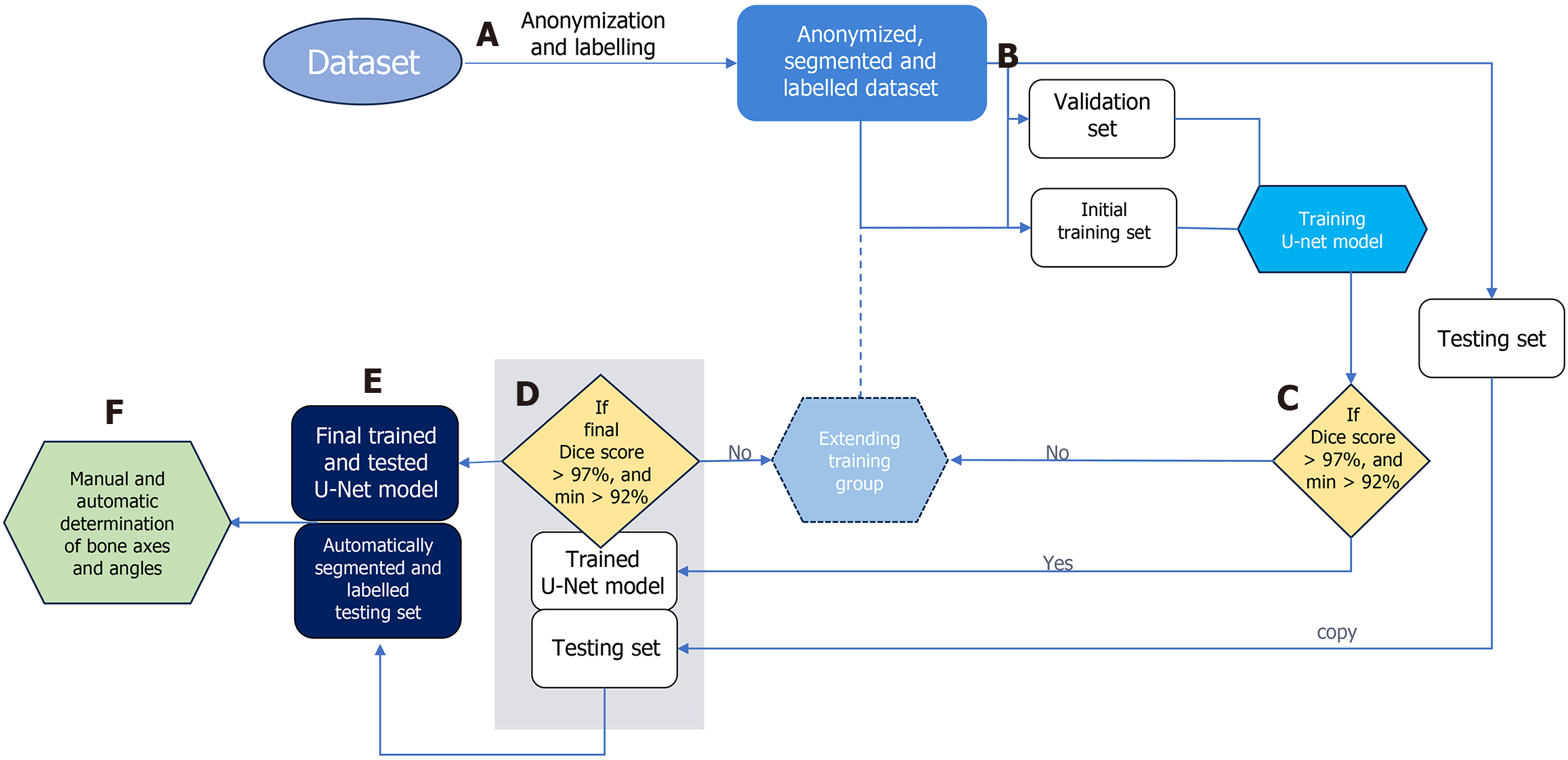
Figure 3 Data flow in the proposed approach.
Stage-A: Bones are manually segmented and labeled from anonymized input radiographs to prepare dataset for multi-class segmentation using a U-Net neural network; Stage-B: Radiographs are randomly divided into three subsets: (1) Training; (2) Validation; and (3) Testing; Stage-C: During each training cycle, the accuracy of bone segmentation by the U-Net is validated on a fixed validation subset of 20 radiographs. The U-Net is initially trained on a subset of 50 radiographs, which is incrementally increased by 10 per cycle until the average Sørensen–Dice index (SDI) exceeds 97% on the validation set; Stage-D: Once the U-Net achieves an SDI > 0.97 on the testing subset, the training process is considered complete. If the SDI does not exceed 0.97, the training subset is expanded, and the network is retrained; Stage-E: The final trained U-Net is used to segment and label bones on all testing radiographs; Stage-F: These segmented bones are then utilized to automatically determine reference points and calculate interphalangeal angle measurements.
- Citation: Kwolek K, Gądek A, Kwolek K, Lechowska-Liszka A, Malczak M, Liszka H. Artificial intelligence-based diagnosis of hallux valgus interphalangeus using anteroposterior foot radiographs. World J Orthop 2025; 16(6): 103832
- URL: https://www.wjgnet.com/2218-5836/full/v16/i6/103832.htm
- DOI: https://dx.doi.org/10.5312/wjo.v16.i6.103832


