Copyright
©The Author(s) 2025.
World J Orthop. May 18, 2025; 16(5): 106377
Published online May 18, 2025. doi: 10.5312/wjo.v16.i5.106377
Published online May 18, 2025. doi: 10.5312/wjo.v16.i5.106377
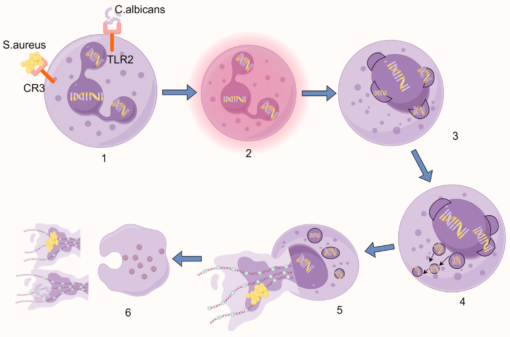
Figure 2 Schematic diagram of vital NETosis.
1: Pathogen recognition via surface receptors triggers cellular activation; 2: Nuclear lobulation is lost, accompanied by chromatin decondensation; 3: The nuclear envelope undergoes sequential disassembly - outer and inner membranes separate and vesiculate; 4: Nuclear-derived vesicles containing stretched chromatin filaments accumulate in the cytoplasm; 5: Cytoplasmic granules migrate toward the intact plasma membrane while maintaining membrane integrity; 6: Focal plasma membrane rupture enables extracellular DNA release, while selective granule exocytosis deposits antimicrobial proteins onto the expelled chromatin network. Created by Figdraw, ID: USWOWe861a. C. albicans: Candida albicans; S. aureus: Staphylococcus aureus; CR3: Complement receptor 3; TLR2: Toll-like receptor 2.
- Citation: Sun GJ, Xu F, Jiao XY, Yin Y. Advances in research of neutrophil extracellular trap formation in osteoarticular diseases. World J Orthop 2025; 16(5): 106377
- URL: https://www.wjgnet.com/2218-5836/full/v16/i5/106377.htm
- DOI: https://dx.doi.org/10.5312/wjo.v16.i5.106377


