Copyright
©The Author(s) 2025.
World J Orthop. May 18, 2025; 16(5): 102916
Published online May 18, 2025. doi: 10.5312/wjo.v16.i5.102916
Published online May 18, 2025. doi: 10.5312/wjo.v16.i5.102916
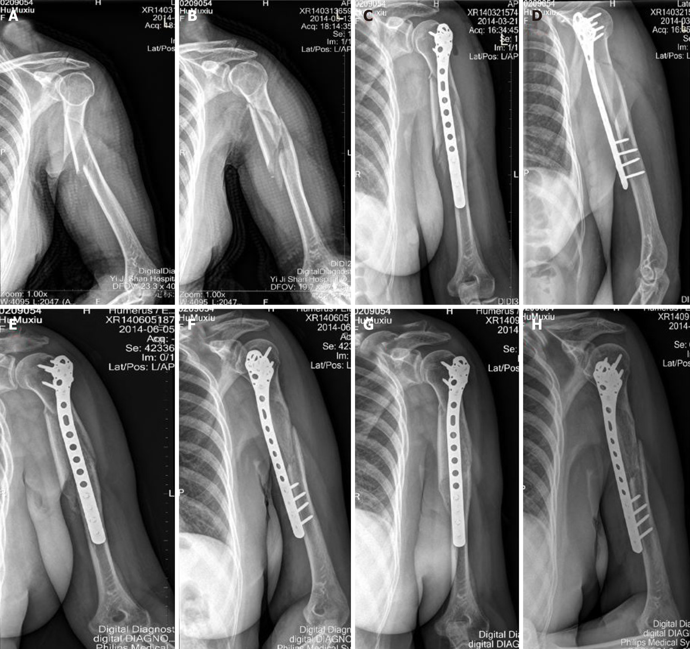
Figure 2 Preoperative, intraoperative, and postoperative X-ray images.
A: Anteroposterior view of preoperative fluoroscopy; B: Lateral radiograph taken before the operation; C: Anteroposterior view during intraoperative fluoroscopy; D: Lateral fluoroscopy during the operation; E: Postoperative anteroposterior radiograph; F: Lateral radiograph after surgery; G: Postoperative multi-angle perspective; H: Postoperative multi-angle perspective again.
- Citation: Cheng WJ, Lu JS, Tao ZS, Xie JB, Yang M. Parallax-free panoramic X-ray imaging combined with minimally invasive plate osteosynthesis for treating proximal humeral shaft fractures. World J Orthop 2025; 16(5): 102916
- URL: https://www.wjgnet.com/2218-5836/full/v16/i5/102916.htm
- DOI: https://dx.doi.org/10.5312/wjo.v16.i5.102916


