Copyright
©The Author(s) 2025.
World J Orthop. Apr 18, 2025; 16(4): 102506
Published online Apr 18, 2025. doi: 10.5312/wjo.v16.i4.102506
Published online Apr 18, 2025. doi: 10.5312/wjo.v16.i4.102506
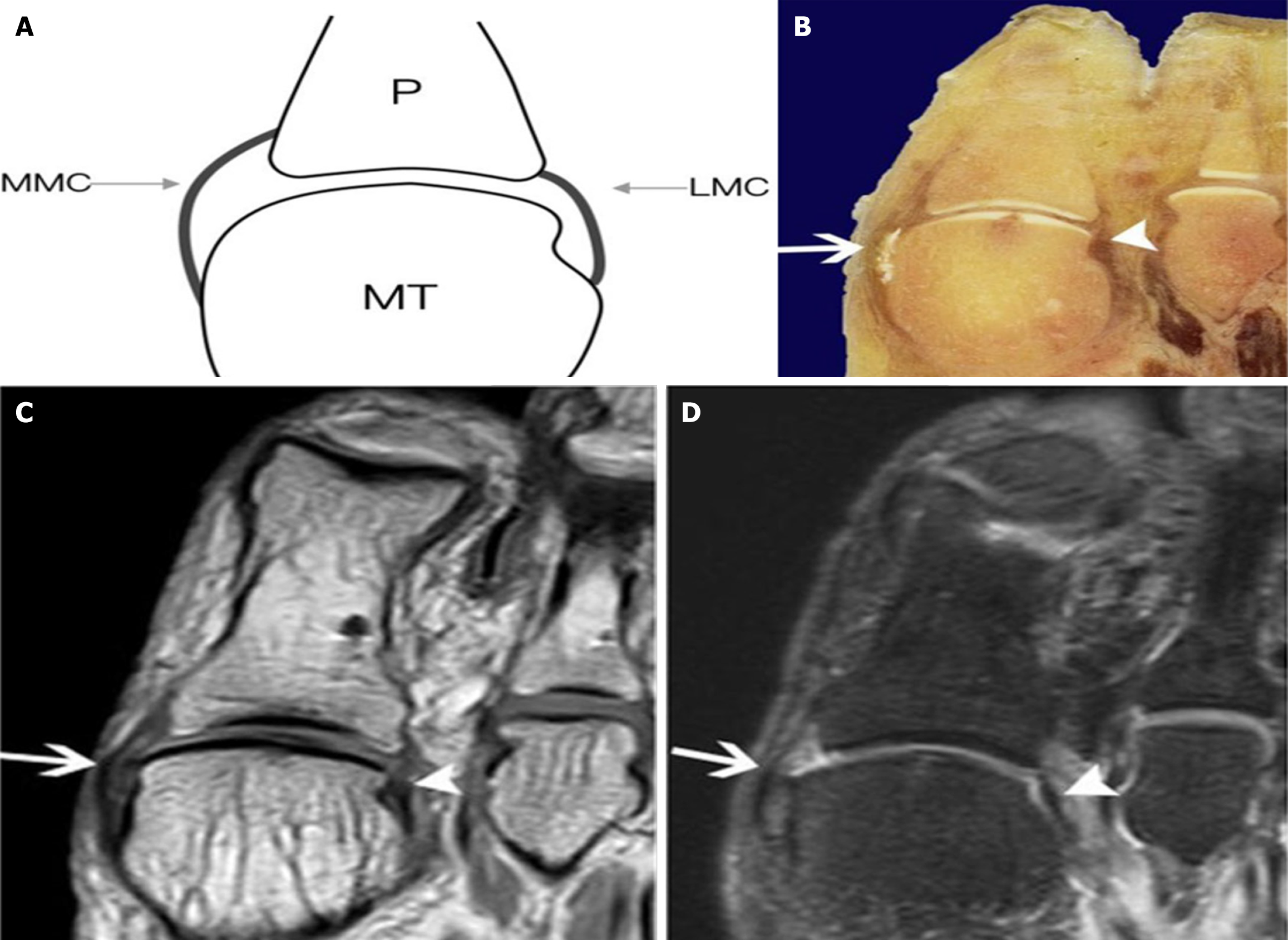
Figure 1 A 33-year-old right foot specimen demonstrating the main lateral and medial collateral ligaments[22].
Citation: Wang JE, Bai RJ, Zhan HL, Li WT, Qian ZH, Wang NL, Yin Y. High-resolution 3T magnetic resonance imaging and histological analysis of capsuloligamentous complex of the first metatarsophalangeal joint. J Orthop Surg Res 2021; 16: 638. Copyright© The Author(s) 2021. Published by Springer Nature. This image is licensed under a Creative Commons Attribution 4.0 International License, which permits use, sharing, adaptation, distribution, and reproduction in any medium, provided the original author(s) and source are credited. License details: https://creativecommons.org/Licenses/by/4.0/. A: Diagrammatic illustration of the first metatarsophalangeal joint; B: Transverse anatomical section; C: T1-weighted magnetic resonance imaging of the foot; D: T2-weighted selective spectral attenuated inversion recovery magnetic resonance imaging of the foot. LMC: Main lateral collateral ligament (white arrowhead); MMC: Main medial collateral ligament (white arrow); MT: Metatarsal; P: Phalanx.
- Citation: Embaby OM, Elalfy MM. First metatarsophalangeal joint: Embryology, anatomy and biomechanics. World J Orthop 2025; 16(4): 102506
- URL: https://www.wjgnet.com/2218-5836/full/v16/i4/102506.htm
- DOI: https://dx.doi.org/10.5312/wjo.v16.i4.102506


