
















©The Author(s) 2025.
World J Orthop. Dec 18, 2025; 16(12): 111602
Published online Dec 18, 2025. doi: 10.5312/wjo.v16.i12.111602
Published online Dec 18, 2025. doi: 10.5312/wjo.v16.i12.111602
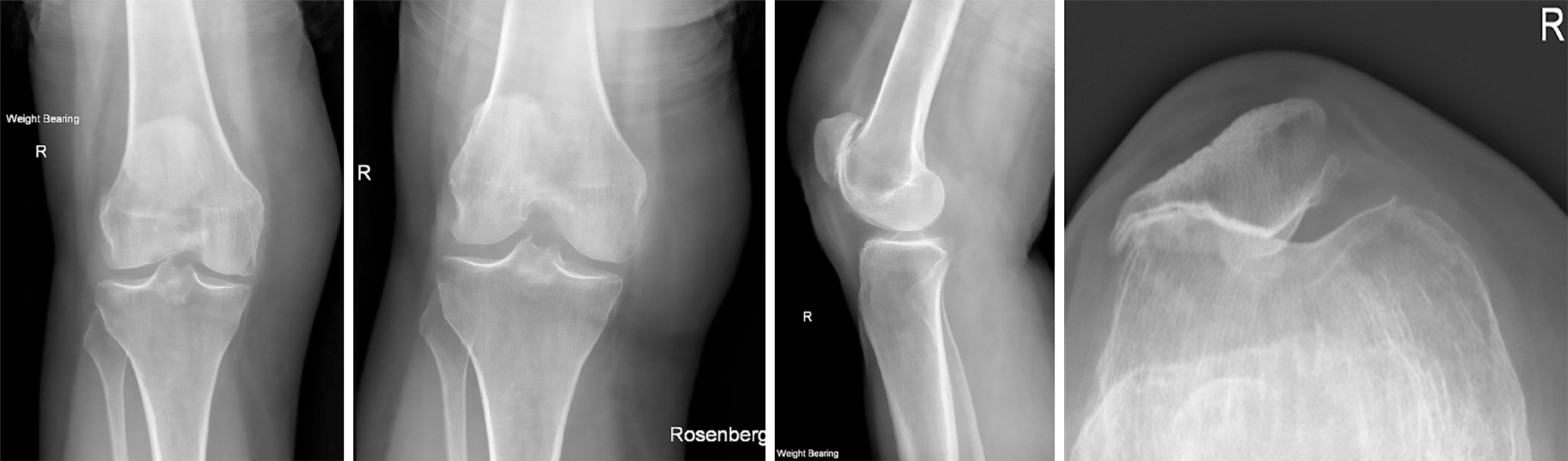
Figure 4 Progress X-rays at 9 months: Interval healing of the fracture at the medial margin of the femoral trochlear, where the fracture is not visualised and alignment appears anatomical.
This is best demonstrated on the skyline projection (bottom right). Background advanced patellofemoral compartment degenerative change is present.
- Citation: Brooks JR, Pitman AG, Lucas P, Cawthorne DP, Wade SWT. Inferior patella dislocation in an elderly patient after a fall: A case report. World J Orthop 2025; 16(12): 111602
- URL: https://www.wjgnet.com/2218-5836/full/v16/i12/111602.htm
- DOI: https://dx.doi.org/10.5312/wjo.v16.i12.111602