
















©The Author(s) 2025.
World J Orthop. Dec 18, 2025; 16(12): 111602
Published online Dec 18, 2025. doi: 10.5312/wjo.v16.i12.111602
Published online Dec 18, 2025. doi: 10.5312/wjo.v16.i12.111602
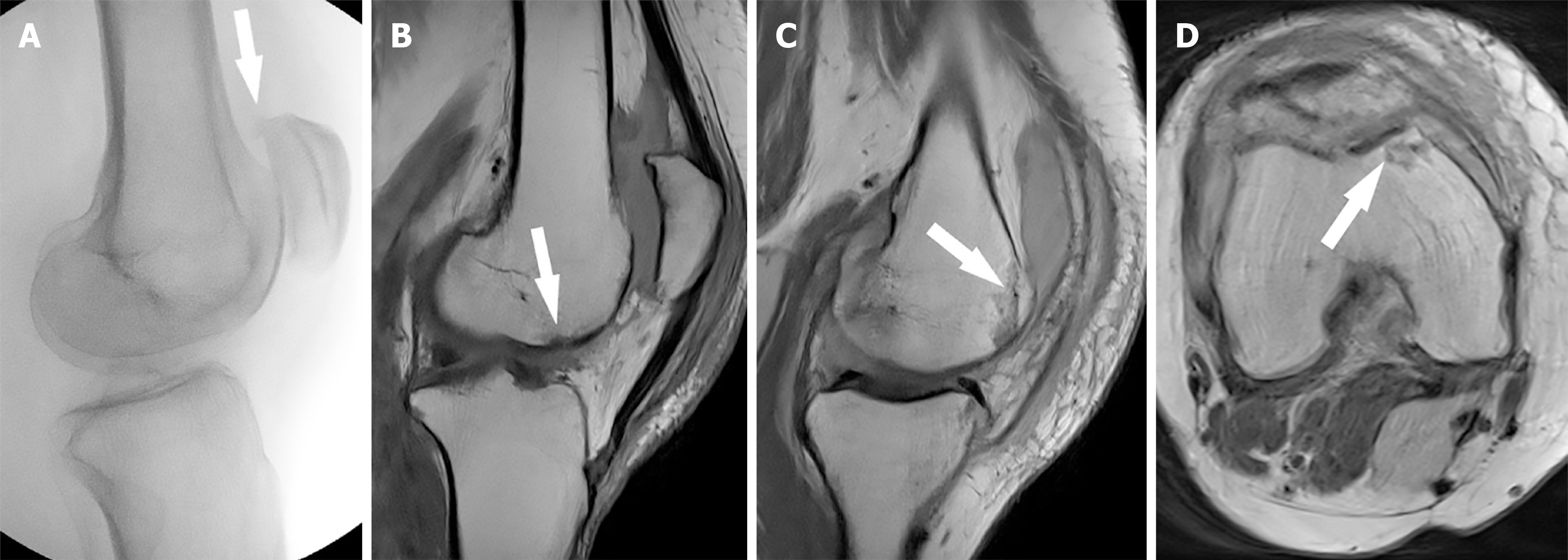
Figure 3 Intraoperative fluoroscopy and postoperative magnetic resonance imaging.
A: Intraoperative fluoroscopy confirmed reduction and showed a small superior patella osteophyte (white arrow); B: The sagittal postoperative magnetic resonance imaging (MRI) sequence redemonstrates the small femoral trochlear osteochondral fracture (white arrow) and confirms an intact extensor mechanism; C: Sagittal MRI sequence demonstrating a nondisplaced osteochondral fracture (white arrow); D: Axial MRI sequence demonstrating a nondisplaced osteochondral fracture (white arrow).
- Citation: Brooks JR, Pitman AG, Lucas P, Cawthorne DP, Wade SWT. Inferior patella dislocation in an elderly patient after a fall: A case report. World J Orthop 2025; 16(12): 111602
- URL: https://www.wjgnet.com/2218-5836/full/v16/i12/111602.htm
- DOI: https://dx.doi.org/10.5312/wjo.v16.i12.111602