
















©The Author(s) 2025.
World J Orthop. Dec 18, 2025; 16(12): 111602
Published online Dec 18, 2025. doi: 10.5312/wjo.v16.i12.111602
Published online Dec 18, 2025. doi: 10.5312/wjo.v16.i12.111602
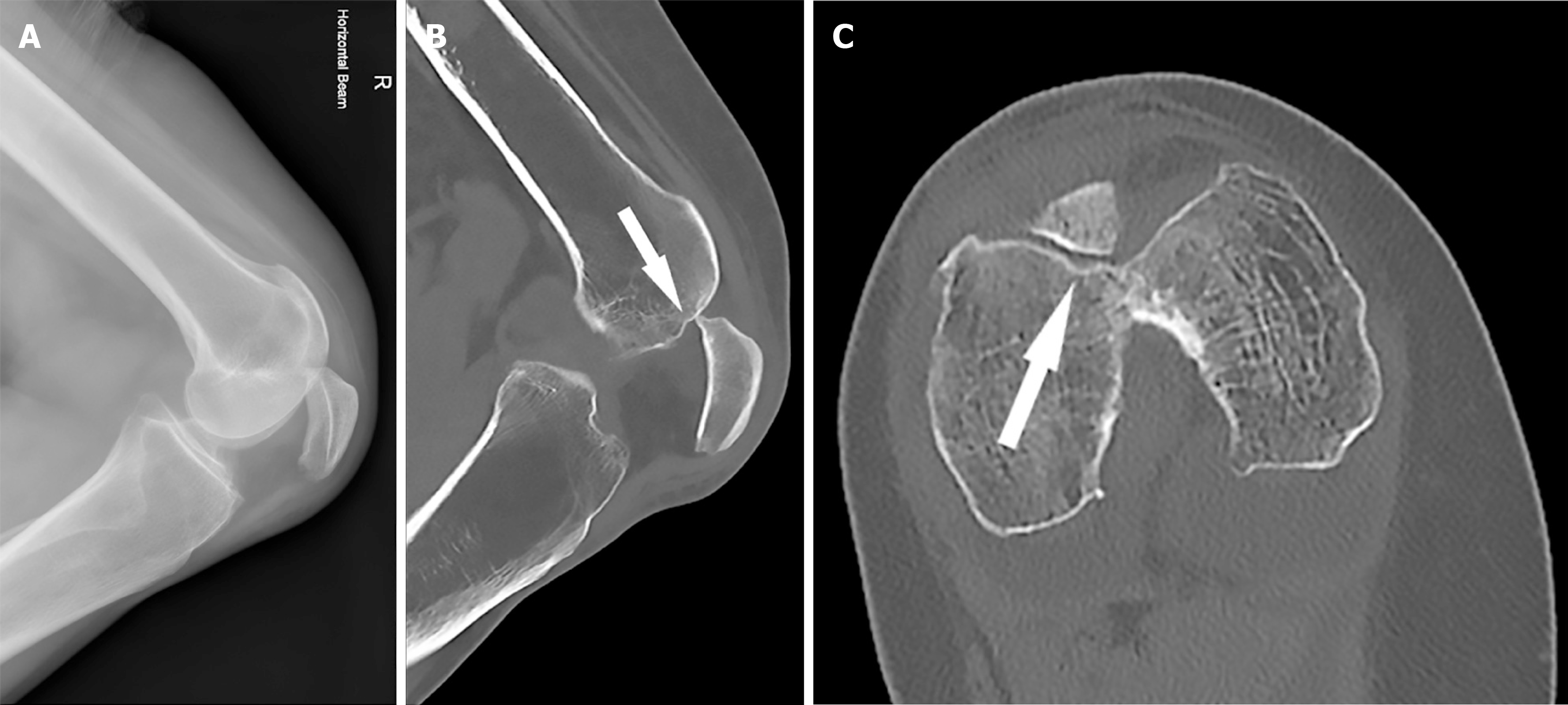
Figure 1 Initial X-ray and computed tomography.
A: Lateral knee X-ray demonstrating the inferior patella dislocation; B: Sagittal right knee computed tomography reconstruction showing a small depressed femoral trochlear fracture (white arrow) with an unchanged patella position; C: Axial right knee computed tomography reconstruction showing the mildly depressed femoral trochlear fracture (white arrow).
- Citation: Brooks JR, Pitman AG, Lucas P, Cawthorne DP, Wade SWT. Inferior patella dislocation in an elderly patient after a fall: A case report. World J Orthop 2025; 16(12): 111602
- URL: https://www.wjgnet.com/2218-5836/full/v16/i12/111602.htm
- DOI: https://dx.doi.org/10.5312/wjo.v16.i12.111602