
















©The Author(s) 2025.
World J Orthop. Dec 18, 2025; 16(12): 111460
Published online Dec 18, 2025. doi: 10.5312/wjo.v16.i12.111460
Published online Dec 18, 2025. doi: 10.5312/wjo.v16.i12.111460
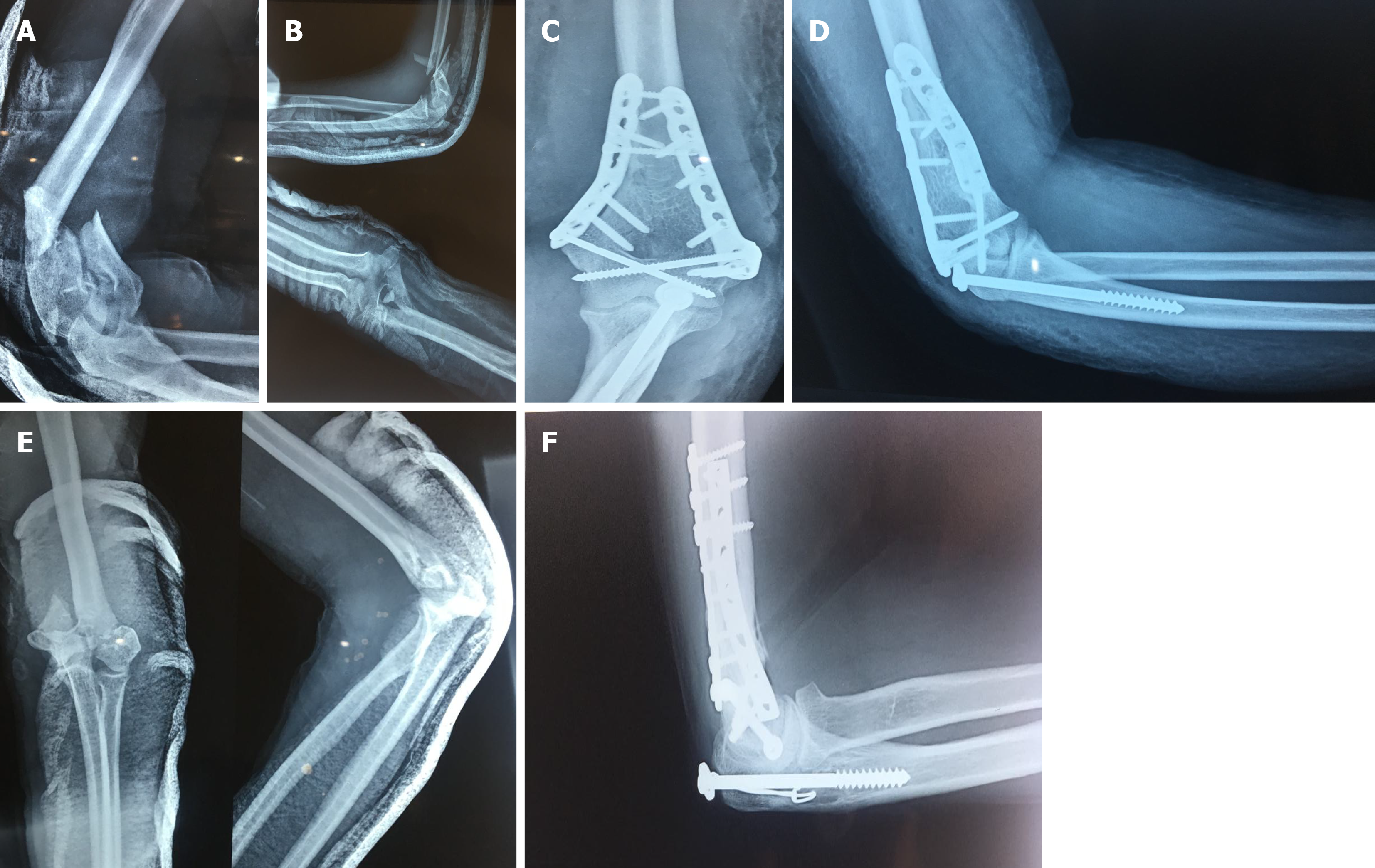
Figure 1 Preoperative and postoperative X-ray films of distal humerus fractures and images of olecranon osteotomy fixation.
A and B: Preoperative anteroposterior (A) and lateral radiographs (B) showing intra-articular distal humerus fracture; C and D: Postoperative anteroposterior (C) and lateral radiographs (D) showing healed fracture distal humerus and healed olecranon osteotomy fixed with cancellous screw and washer; E and F: Preoperative and postoperative radiographs showing fixation of olecranon osteotomy with intramedullary screw and cerclage. Preoperative anteroposterior and lateral radiographs showing intra-articular distal humerus fracture (E). Fixation with pre-contoured locking plates and olecranon osteotomy fixation with intramedullary screw and cerclage (F).
- Citation: Shah FA, Sattar A, Iqbal J. Comparison of two fixation techniques of olecranon osteotomy after reconstruction of intra-articular distal humerus fractures. World J Orthop 2025; 16(12): 111460
- URL: https://www.wjgnet.com/2218-5836/full/v16/i12/111460.htm
- DOI: https://dx.doi.org/10.5312/wjo.v16.i12.111460