
















©The Author(s) 2025.
World J Orthop. Nov 18, 2025; 16(11): 112307
Published online Nov 18, 2025. doi: 10.5312/wjo.v16.i11.112307
Published online Nov 18, 2025. doi: 10.5312/wjo.v16.i11.112307
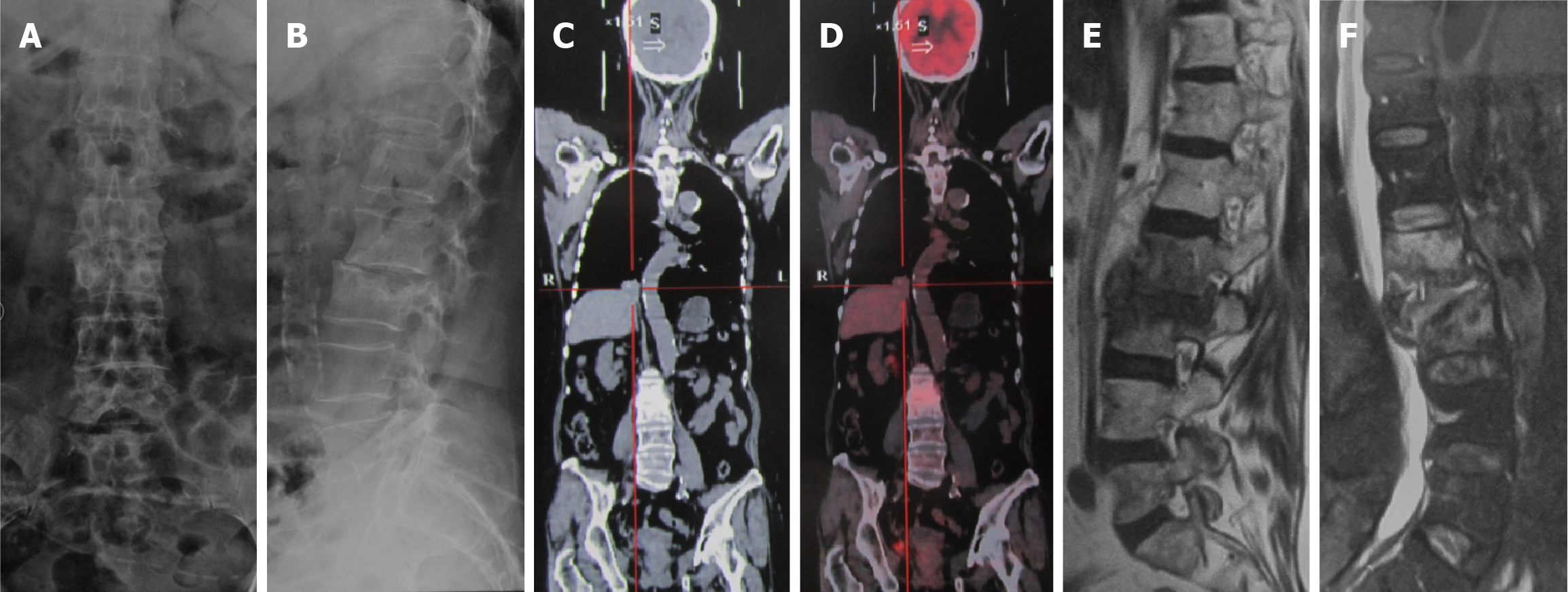
Figure 1 Imaging examinations before surgery.
A and B: Visible destruction of the L2 and L3 vertebral bodies, with narrowing of the L2/L3 space; C and D: Positron emission tomography/computed tomography suggest bone metastasis of lung cancer to the L2 and L3 vertebrae; E and F: Magnetic resonance imaging indicates increased signals in the L2 and L3 vertebral bodies and intervertebral space.
- Citation: Pan JL, Xing H, Li Y, Chang ZQ. Successful treatment of a non-healing lumbar incision post-radiotherapy using vacuum sealing drainage: A case report. World J Orthop 2025; 16(11): 112307
- URL: https://www.wjgnet.com/2218-5836/full/v16/i11/112307.htm
- DOI: https://dx.doi.org/10.5312/wjo.v16.i11.112307