
















©The Author(s) 2025.
World J Orthop. Nov 18, 2025; 16(11): 111052
Published online Nov 18, 2025. doi: 10.5312/wjo.v16.i11.111052
Published online Nov 18, 2025. doi: 10.5312/wjo.v16.i11.111052
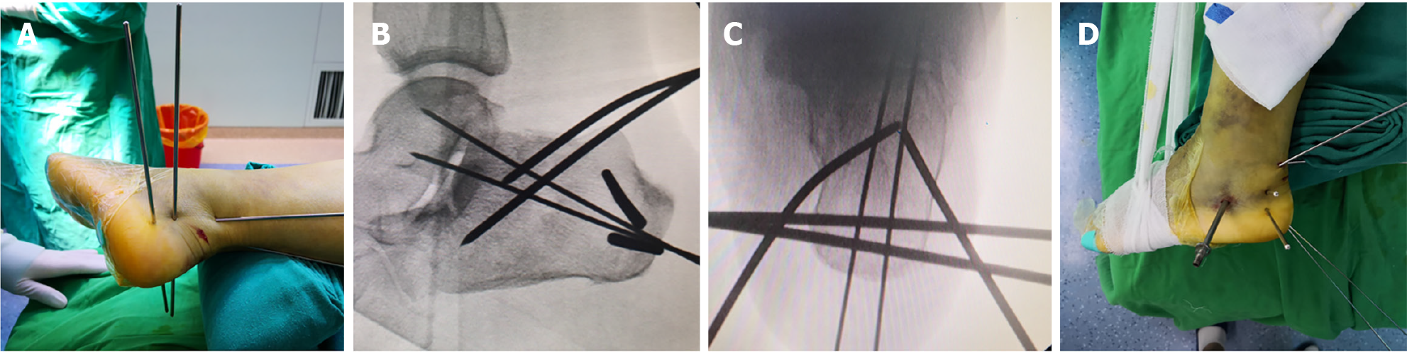
Figure 4 Intraoperative fluoroscopy demonstrating the reduction procedure and outcomes.
A: After prying reduction restored the normal Böhler’s angle, the Steinmann pin was used to engage the distal fracture fragment posterior-inferior to the calcaneal body for eversion reduction; B and C: With compression applied to both sides of the calcaneus, fluoroscopy confirmed satisfactory reduction. A 2.5 mm guide wire was then drilled from the posterior calcaneus in an anterior direction; D: Post-reduction photograph.
- Citation: Huang HC, Che YF, Sun H, Xu YS, Gao HY, Tang XS. Traditional Chinese bone-setting combined with percutaneous screw fixation for comminuted calcaneal fractures: A case report and review of literature. World J Orthop 2025; 16(11): 111052
- URL: https://www.wjgnet.com/2218-5836/full/v16/i11/111052.htm
- DOI: https://dx.doi.org/10.5312/wjo.v16.i11.111052