
















©The Author(s) 2025.
World J Orthop. Nov 18, 2025; 16(11): 111052
Published online Nov 18, 2025. doi: 10.5312/wjo.v16.i11.111052
Published online Nov 18, 2025. doi: 10.5312/wjo.v16.i11.111052
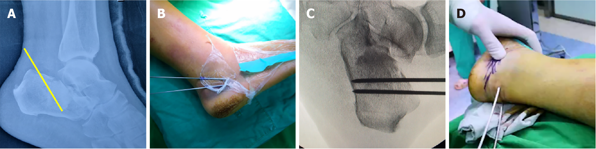
Figure 3 Step 1.
A: The preoperative plan specified a 40° prying angle with the entry point at the anterior margin of the calcaneal tuberosity. The tip of the Steinmann pin was positioned at the deepest point of the depressed fracture fragment; B: Two 2.0 mm Kirschner wires were placed superficially to simulate the intended trajectory and depth of insertion; C: Two 3.0 mm Steinmann pins were then inserted into the calcaneus with approximately 1.5 cm spacing between them, following the trajectory established by the superficial Kirschner wires; D: Fluoroscopic imaging confirmed satisfactory positioning of the superficial Kirschner wires.
- Citation: Huang HC, Che YF, Sun H, Xu YS, Gao HY, Tang XS. Traditional Chinese bone-setting combined with percutaneous screw fixation for comminuted calcaneal fractures: A case report and review of literature. World J Orthop 2025; 16(11): 111052
- URL: https://www.wjgnet.com/2218-5836/full/v16/i11/111052.htm
- DOI: https://dx.doi.org/10.5312/wjo.v16.i11.111052