
















©The Author(s) 2025.
World J Orthop. Nov 18, 2025; 16(11): 110716
Published online Nov 18, 2025. doi: 10.5312/wjo.v16.i11.110716
Published online Nov 18, 2025. doi: 10.5312/wjo.v16.i11.110716
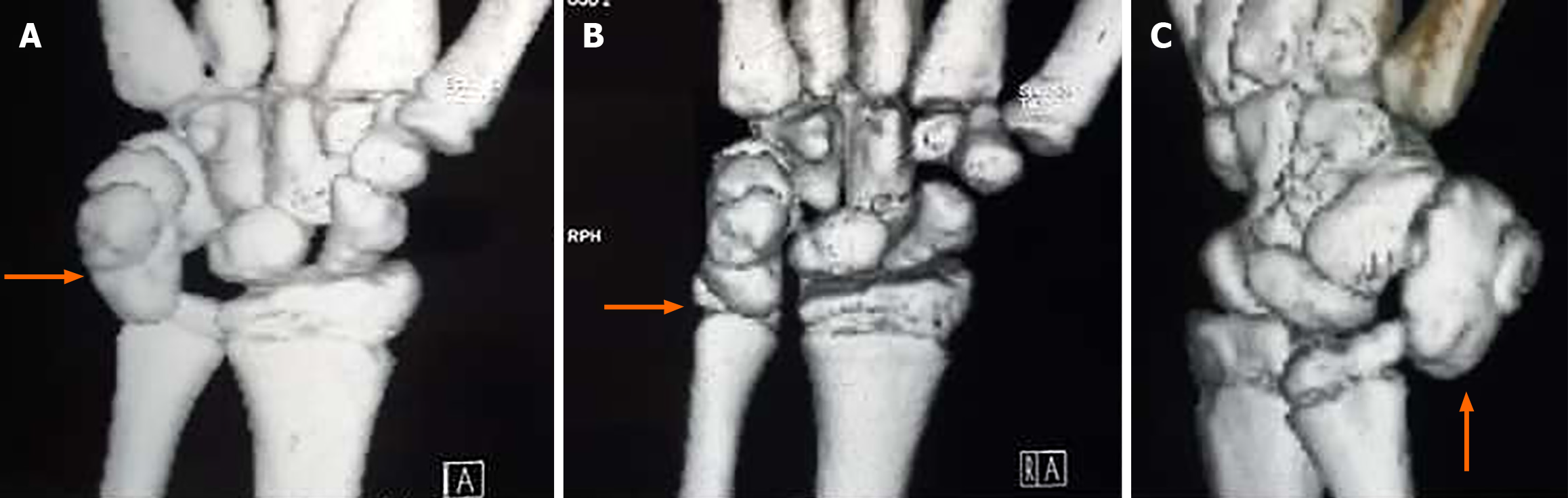
Figure 2 Computed tomography scan demonstrated a well-circumscribed lesion and a small bump ventrally suggestive of pisiform (pisiform with dorsal osteochondroma) with no stalk and no periosteal reaction (3D computed tomography marked).
A-C: Various views of the wrist (anteroposterior and oblique) and the orange arrow pointing the lesion.
- Citation: Vanapalli R, Jayant UK, Mlv SK, Rawat SS, Ansari MT. Osteochondroma of the pisiform: A case report. World J Orthop 2025; 16(11): 110716
- URL: https://www.wjgnet.com/2218-5836/full/v16/i11/110716.htm
- DOI: https://dx.doi.org/10.5312/wjo.v16.i11.110716