
















©The Author(s) 2025.
World J Orthop. Nov 18, 2025; 16(11): 110426
Published online Nov 18, 2025. doi: 10.5312/wjo.v16.i11.110426
Published online Nov 18, 2025. doi: 10.5312/wjo.v16.i11.110426
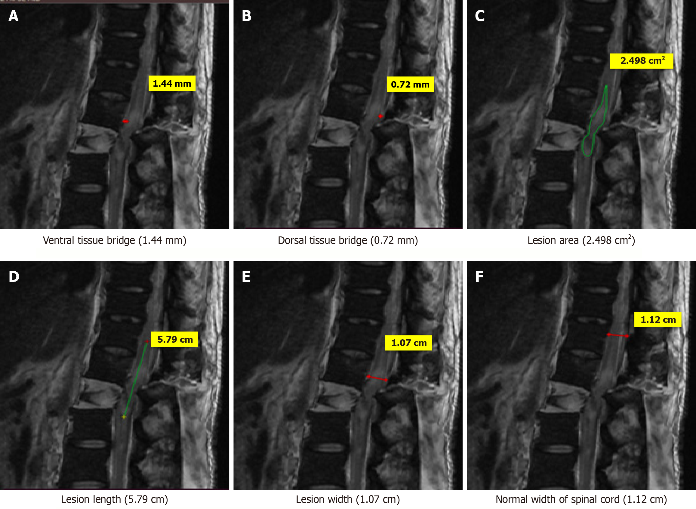
Figure 5 Magnetic resonance imaging T2-weighted sagittal image of a 34-years old patient who sustained trauma of thoracolumbar spine (D12-L1) with ASIA grade A.
A: Ventral tissue bridge (1.44 mm); B: Dorsal tissue bridge (0.72 mm); C: Lesion area (2.498 cm2); D: Lesion length (5.79 cm); E: Lesion width (1.07 cm); F: Normal width of spinal cord (1.12 cm).
- Citation: Singh R, Gautam S, Aggarwal S, Kaur S, Jain M. Correlation of magnetic resonance imaging biomarkers (tissue bridges) with neurological recovery following traumatic spinal cord injury. World J Orthop 2025; 16(11): 110426
- URL: https://www.wjgnet.com/2218-5836/full/v16/i11/110426.htm
- DOI: https://dx.doi.org/10.5312/wjo.v16.i11.110426