
















©The Author(s) 2025.
World J Orthop. Nov 18, 2025; 16(11): 110173
Published online Nov 18, 2025. doi: 10.5312/wjo.v16.i11.110173
Published online Nov 18, 2025. doi: 10.5312/wjo.v16.i11.110173
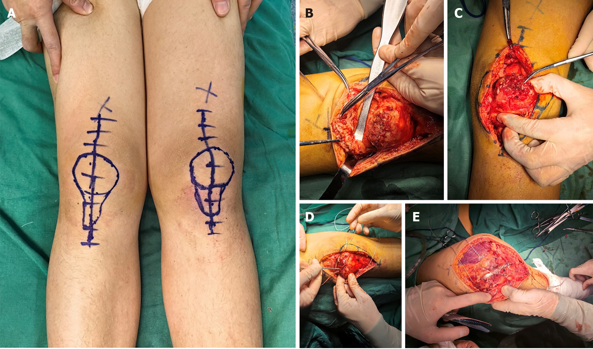
Figure 3 Preoperative and intraoperative photos.
A: Patient in the supine position with an anterior approach incision in both knees; B-E: Intraoperative photographs. A thin shell of avulsed bone in the right knee (B); A thin shell of avulsed bone in the left knee (C); Two 4.5-mm suture anchors placed on the upper part of the patella in the right knee (D); The autogenous gracilis was applied to augment the stability by performing the figure-of-eight technique in the right knee (E).
- Citation: He WP, Ren CM, Luo F, Chen L, Wang WT, Qiu BT, Zhang XC, Chen HT. Bilateral sleeve fracture of the superior pole of the patella in a healthy adult: A case report. World J Orthop 2025; 16(11): 110173
- URL: https://www.wjgnet.com/2218-5836/full/v16/i11/110173.htm
- DOI: https://dx.doi.org/10.5312/wjo.v16.i11.110173