
















©The Author(s) 2025.
World J Orthop. Nov 18, 2025; 16(11): 110093
Published online Nov 18, 2025. doi: 10.5312/wjo.v16.i11.110093
Published online Nov 18, 2025. doi: 10.5312/wjo.v16.i11.110093
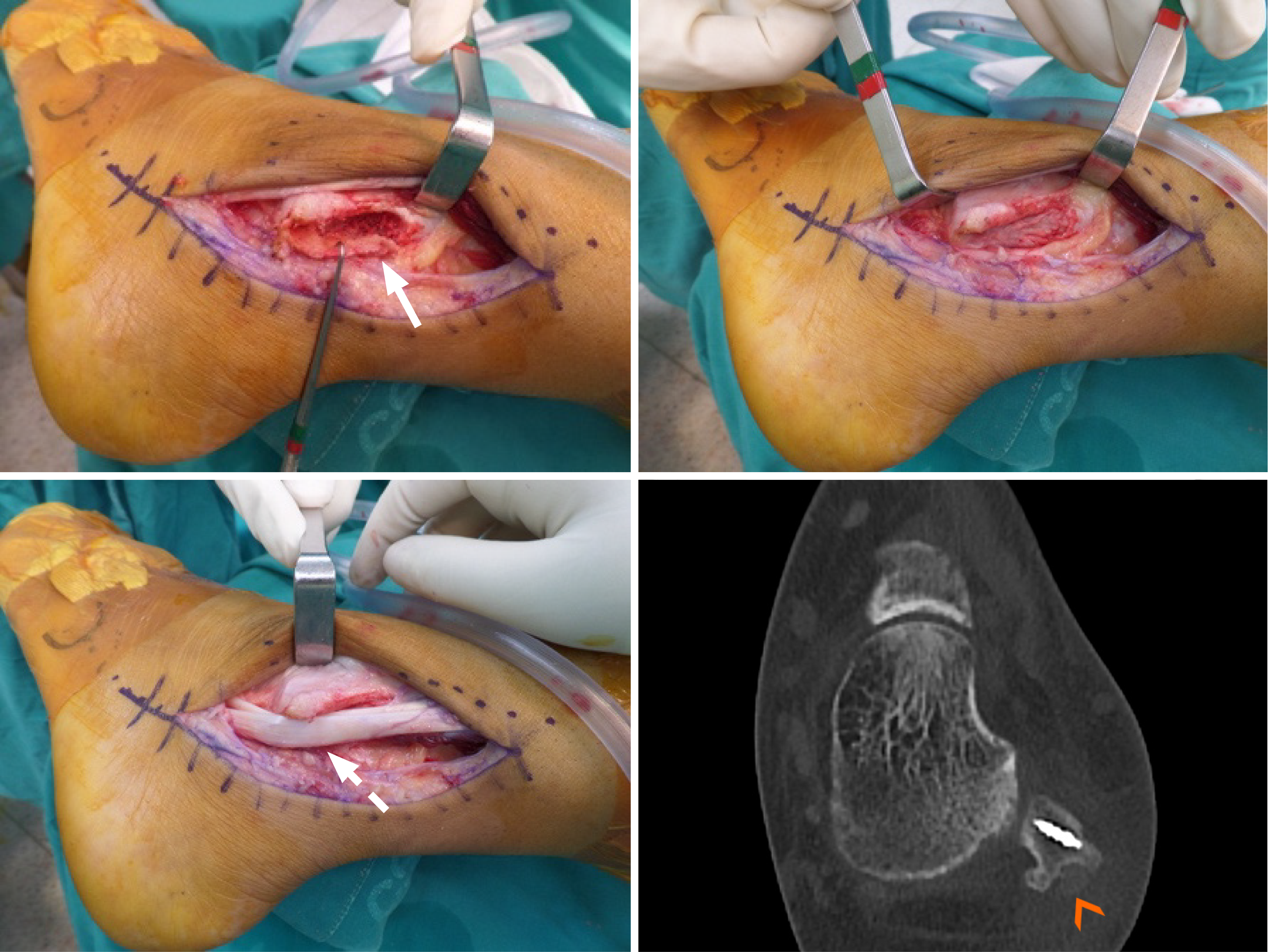
Figure 6 Intraoperative images of a groove deepening procedure: Creating an osteocartilagenous flap (arrow), impacting the underlying cancellous bone to deepen the groove before repositioning the peroneal tendons (dotted arrow).
A postoperative computed tomography image demonstrates the recreated peroneal groove (arrowhead) and superior peroneal retinaculum repair using a suture anchor.
- Citation: Riewruja K, Wattanatanagorn T, Apinun J. Snapping phenomenon around the ankle: An anatomy-based review. World J Orthop 2025; 16(11): 110093
- URL: https://www.wjgnet.com/2218-5836/full/v16/i11/110093.htm
- DOI: https://dx.doi.org/10.5312/wjo.v16.i11.110093