
















©The Author(s) 2025.
World J Orthop. Oct 18, 2025; 16(10): 108992
Published online Oct 18, 2025. doi: 10.5312/wjo.v16.i10.108992
Published online Oct 18, 2025. doi: 10.5312/wjo.v16.i10.108992
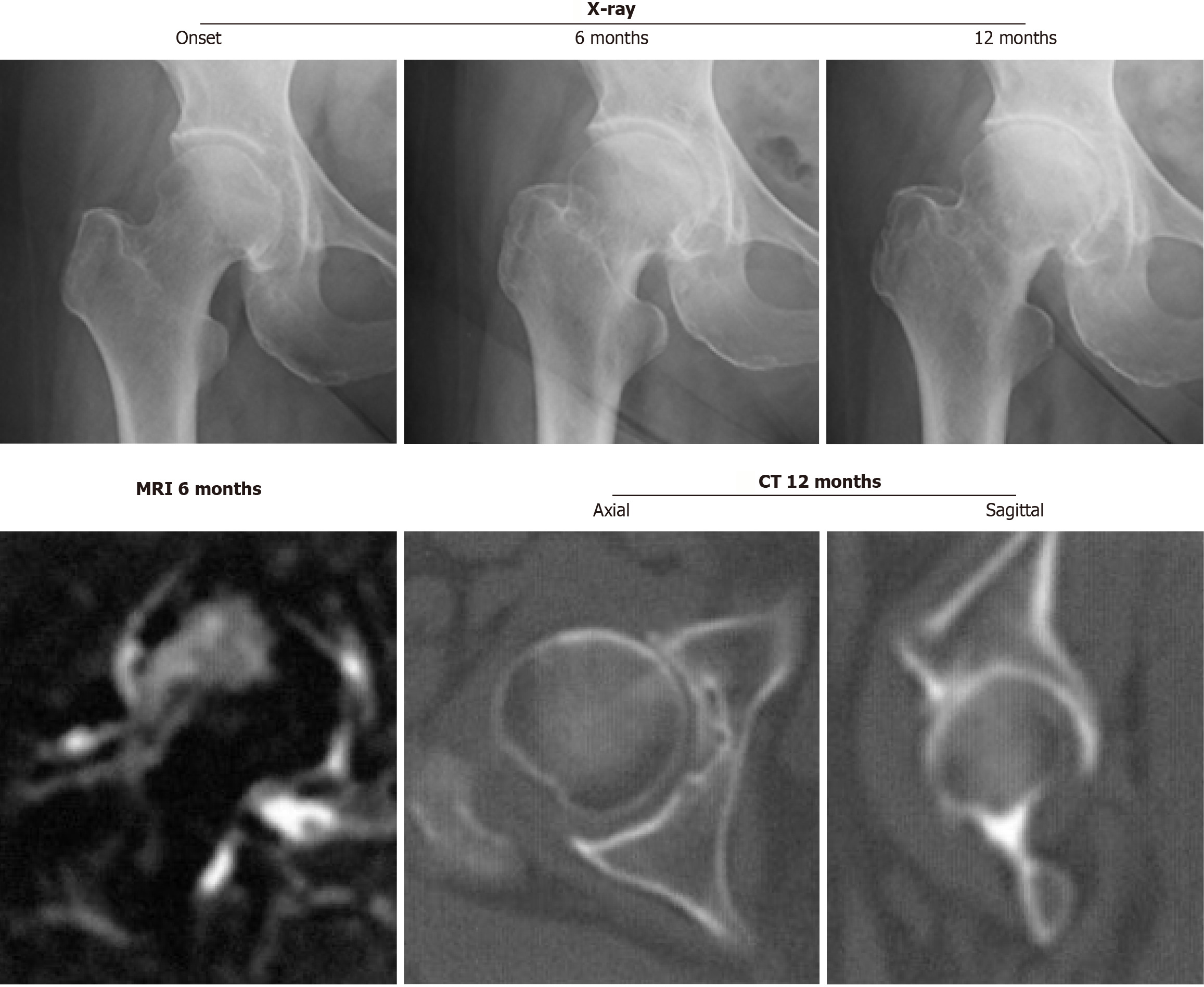
Figure 1 Right hip joint with rapidly progressive osteoarthritis of the hip showing chondrolysis greater than 2 mm/year on a series of radiographs without femoral head destruction on computed tomography at 12 months after the onset.
Magnetic resonance imaging at 6 months after the onset demonstrates inhomogeneous high intensity on the short τ inversion recovery sequence image, including the superolateral portion of the femoral head. CT: Computed tomography; MRI: Magnetic resonance imaging.
- Citation: Yasuda T. Early diagnosis and targeted intervention based on the pathogenesis of rapidly progressive osteoarthritis of the hip. World J Orthop 2025; 16(10): 108992
- URL: https://www.wjgnet.com/2218-5836/full/v16/i10/108992.htm
- DOI: https://dx.doi.org/10.5312/wjo.v16.i10.108992