
















©The Author(s) 2024.
World J Orthop. Sep 18, 2024; 15(9): 891-901
Published online Sep 18, 2024. doi: 10.5312/wjo.v15.i9.891
Published online Sep 18, 2024. doi: 10.5312/wjo.v15.i9.891
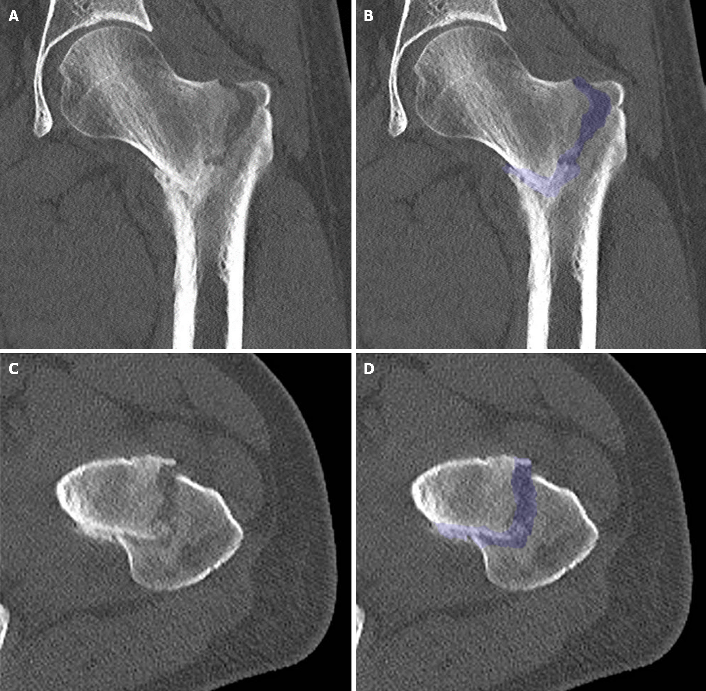
Figure 3 Computed tomography scan at presentation.
A: Coronal section without overlay; B: Coronal section including an overlay, highlighting the fracture; C: Transverse section without overlay; D: Transverse section with an overlay, highlighting the fracture. Additional imaging with computed tomography did not reveal evidence of pre-existing bone pathology, but confirmed significant impaction and malalignment. Furthermore, there were multiple signs of osteopenia in the left leg, most likely due to disuse over the preceding 3 weeks. Additionally, there was thickening of the periosteum on the medial side halfway down the femur, possibly due to altered force distribution. In the transverse section, external rotation is notably observed.
- Citation: Oudmaijer CA, Paulino Pereira NR, Visser D, Wakker AM, Veltman ES, van Linschoten R. Lateral femoral neck stress fractures: A case report. World J Orthop 2024; 15(9): 891-901
- URL: https://www.wjgnet.com/2218-5836/full/v15/i9/891.htm
- DOI: https://dx.doi.org/10.5312/wjo.v15.i9.891