Copyright
©The Author(s) 2022.
World J Orthop. May 18, 2022; 13(5): 528-537
Published online May 18, 2022. doi: 10.5312/wjo.v13.i5.528
Published online May 18, 2022. doi: 10.5312/wjo.v13.i5.528
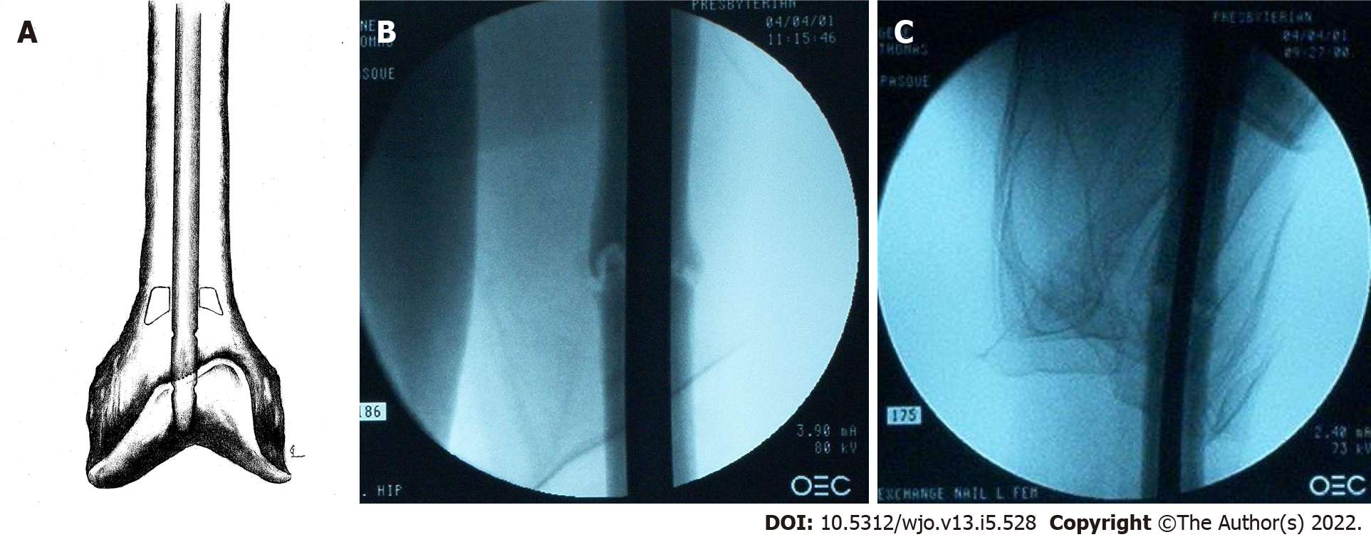
Figure 6 Illustration and fluoroscopic radiographs of left femur obtained intra-operatively.
A: Illustration showing intramedullary nail placed past bone pedestal; B: Anterior-posterior radiograph showing new, larger diameter intramedullary nail placed past bone pedestal; C: Lateral radiograph showing the same.
- Citation: Pasque CB, Pappas AJ, Cole Jr CA. Intramedullary bone pedestal formation contributing to femoral shaft fracture nonunion: A case report and review of the literature. World J Orthop 2022; 13(5): 528-537
- URL: https://www.wjgnet.com/2218-5836/full/v13/i5/528.htm
- DOI: https://dx.doi.org/10.5312/wjo.v13.i5.528


