Copyright
©The Author(s) 2022.
World J Orthop. May 18, 2022; 13(5): 528-537
Published online May 18, 2022. doi: 10.5312/wjo.v13.i5.528
Published online May 18, 2022. doi: 10.5312/wjo.v13.i5.528
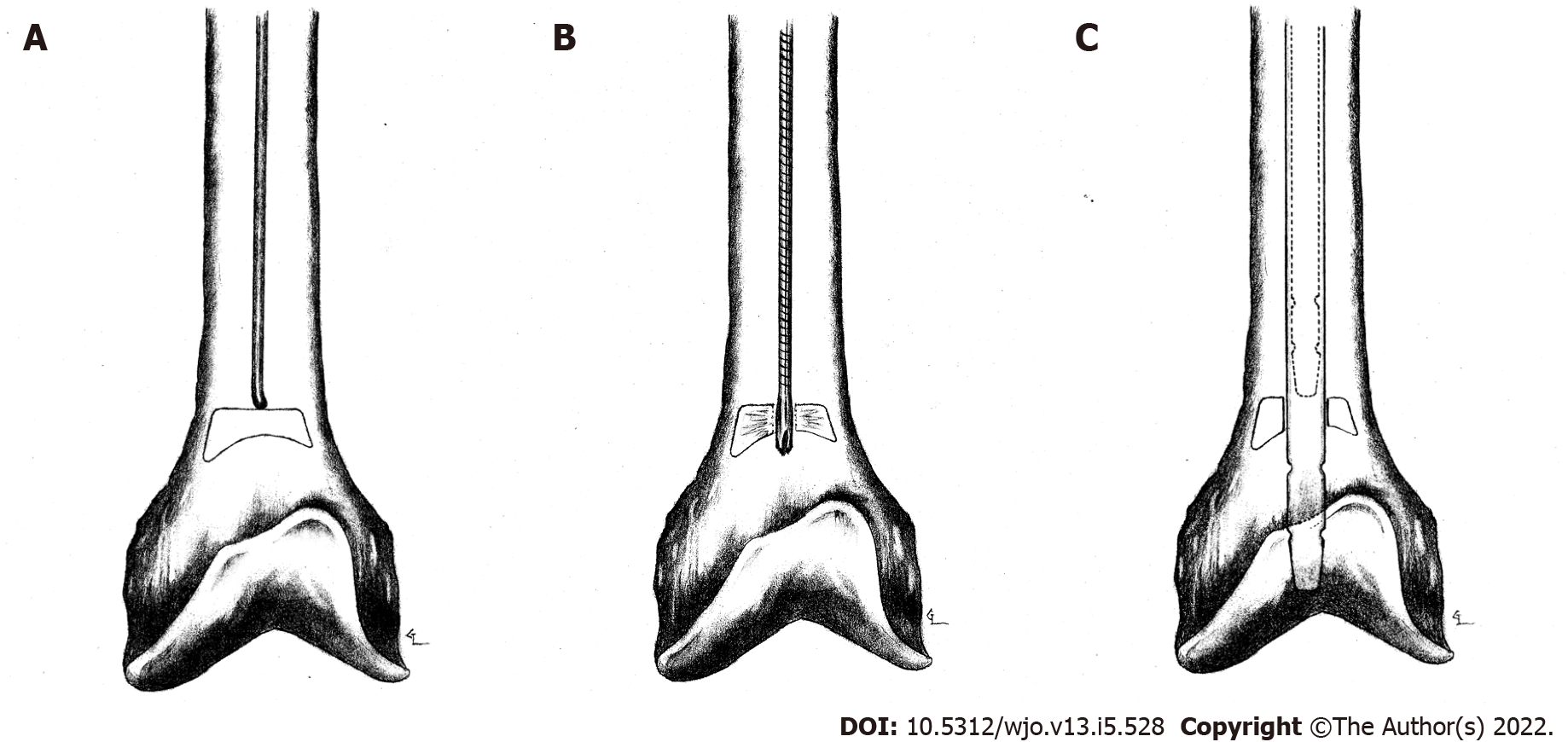
Figure 5 Illustration of left femur intra-operatively.
A: Showing femur after intramedullary nail removal. Guide rod tip still unable to pass distally in canal due to intramedullary bone pedestal; B: Showing starting reamer used to breach intramedullary bone pedestal; C: Showing new nail (solid lines) at area of wider, more distal meta-diaphyseal bone compared to old nail (dotted lines) at area of more proximal, narrow diaphyseal bone.
- Citation: Pasque CB, Pappas AJ, Cole Jr CA. Intramedullary bone pedestal formation contributing to femoral shaft fracture nonunion: A case report and review of the literature. World J Orthop 2022; 13(5): 528-537
- URL: https://www.wjgnet.com/2218-5836/full/v13/i5/528.htm
- DOI: https://dx.doi.org/10.5312/wjo.v13.i5.528


