Copyright
©The Author(s) 2022.
World J Orthop. May 18, 2022; 13(5): 515-527
Published online May 18, 2022. doi: 10.5312/wjo.v13.i5.515
Published online May 18, 2022. doi: 10.5312/wjo.v13.i5.515
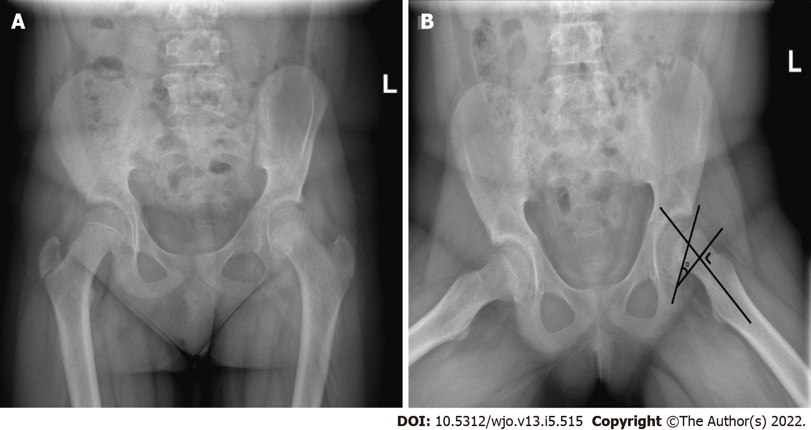
Figure 1 Plain pelvic radiographs of a left-sided slipped capital femoral epiphysis showing the posterior sloping angle of the affected side.
A high posterior sloping angle of the unaffected side is considered an independent risk factor for subsequent contralateral disease. The posterior sloping angle (α), here presented on the affected side to increase visibility, is measured as the angle formed by the line along the physeal plane and the line perpendicular to the femoral neck-diaphyseal axis. A: Anteroposterior view; B: Lauenstein view.
- Citation: Vink SJC, van Stralen RA, Moerman S, van Bergen CJA. Prophylactic fixation of the unaffected contralateral side in children with slipped capital femoral epiphysis seems favorable: A systematic review. World J Orthop 2022; 13(5): 515-527
- URL: https://www.wjgnet.com/2218-5836/full/v13/i5/515.htm
- DOI: https://dx.doi.org/10.5312/wjo.v13.i5.515


