Copyright
©The Author(s) 2021.
World J Orthop. Jun 18, 2021; 12(6): 445-455
Published online Jun 18, 2021. doi: 10.5312/wjo.v12.i6.445
Published online Jun 18, 2021. doi: 10.5312/wjo.v12.i6.445
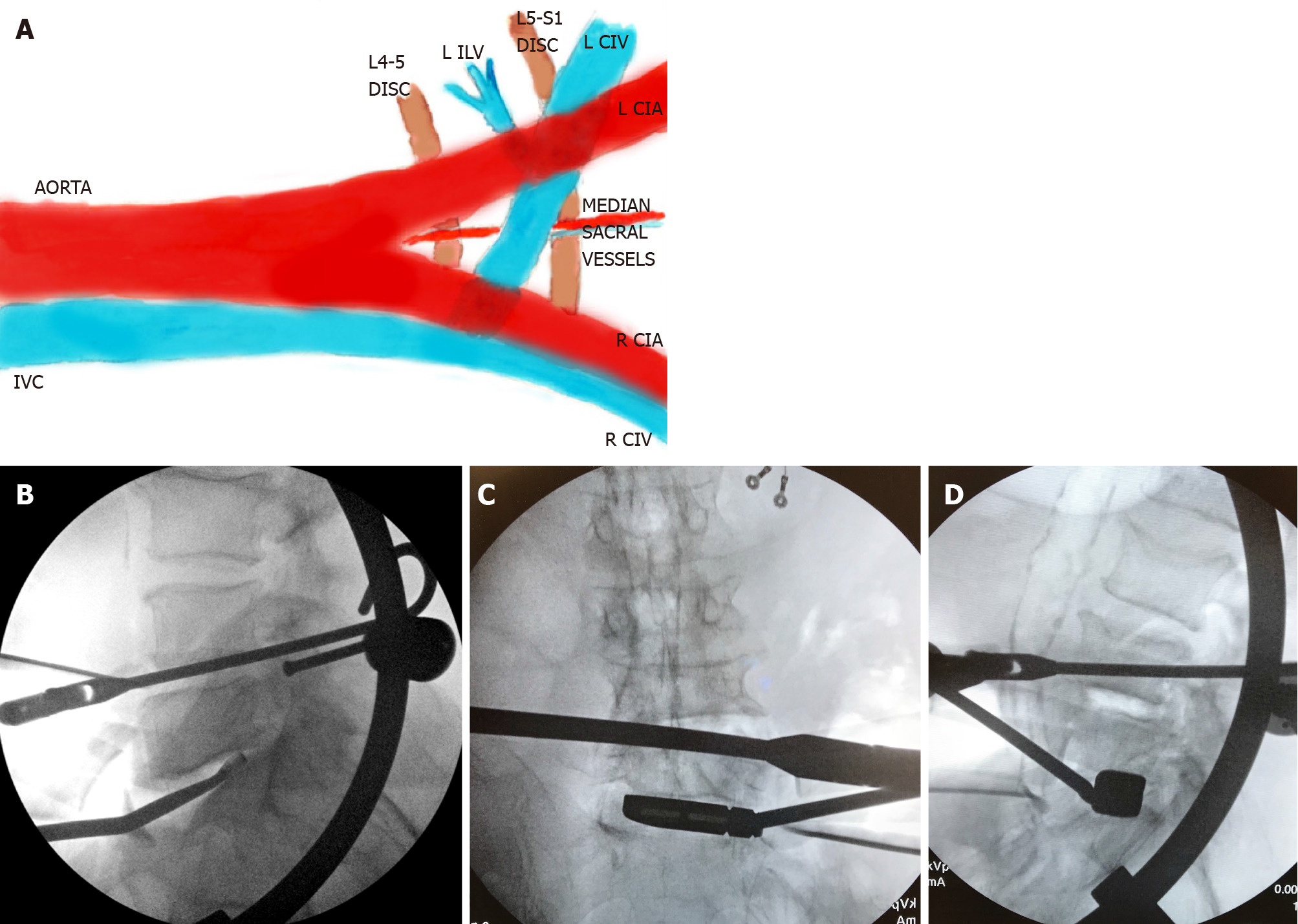
Figure 3 Illustration showing left-sided oblique anterolateral approaches (A), and intraoperative fluoroscopy images (B-D).
Image A shows the median sacral vessels that are often encountered in the left intra-bifurcation approach, and the left ilio-lumbar vein (ILV), which may need ligation in a left pre-psoas approach. Image B shows a lateral fluoroscopy image with a double-bent curette which may be helpful in preparing the portions of the L5 inferior endplate that may not be under direct visualization. Images C and D show intraoperative anteroposterior and lateral fluoroscopy images during a left pre-psoas approach with specialized trial instrument bent in two planes. IVC: Inferior vena cava; CIV: Common iliac vein; CIA: Common iliac artery; ILV: Ilio-lumbar vein; R: Right; L: Left.
- Citation: Berry CA. Nuances of oblique lumbar interbody fusion at L5-S1: Three case reports. World J Orthop 2021; 12(6): 445-455
- URL: https://www.wjgnet.com/2218-5836/full/v12/i6/445.htm
- DOI: https://dx.doi.org/10.5312/wjo.v12.i6.445


