Copyright
©The Author(s) 2021.
World J Orthop. May 18, 2021; 12(5): 329-337
Published online May 18, 2021. doi: 10.5312/wjo.v12.i5.329
Published online May 18, 2021. doi: 10.5312/wjo.v12.i5.329
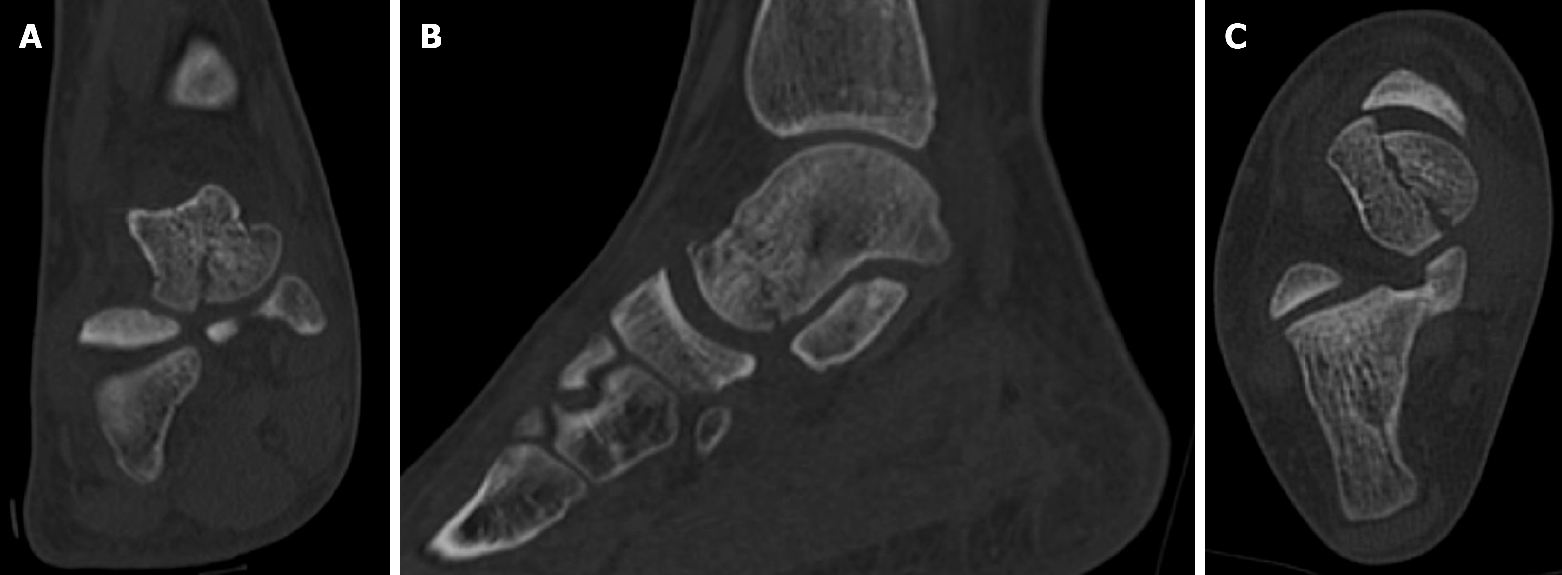
Figure 2 Pre-operative computed tomography images of the shear-type fracture.
A: Coronal, B: Sagittal; and C: Axial view are shown.
- Citation: Monestier L, Riva G, Faoro L, Surace MF. Rare shear-type fracture of the talar head in a thirteen-year-old child — Is this a transitional fracture: A case report and review of the literature. World J Orthop 2021; 12(5): 329-337
- URL: https://www.wjgnet.com/2218-5836/full/v12/i5/329.htm
- DOI: https://dx.doi.org/10.5312/wjo.v12.i5.329


