Copyright
©The Author(s) 2020.
World J Orthop. Mar 18, 2020; 11(3): 145-157
Published online Mar 18, 2020. doi: 10.5312/wjo.v11.i3.145
Published online Mar 18, 2020. doi: 10.5312/wjo.v11.i3.145
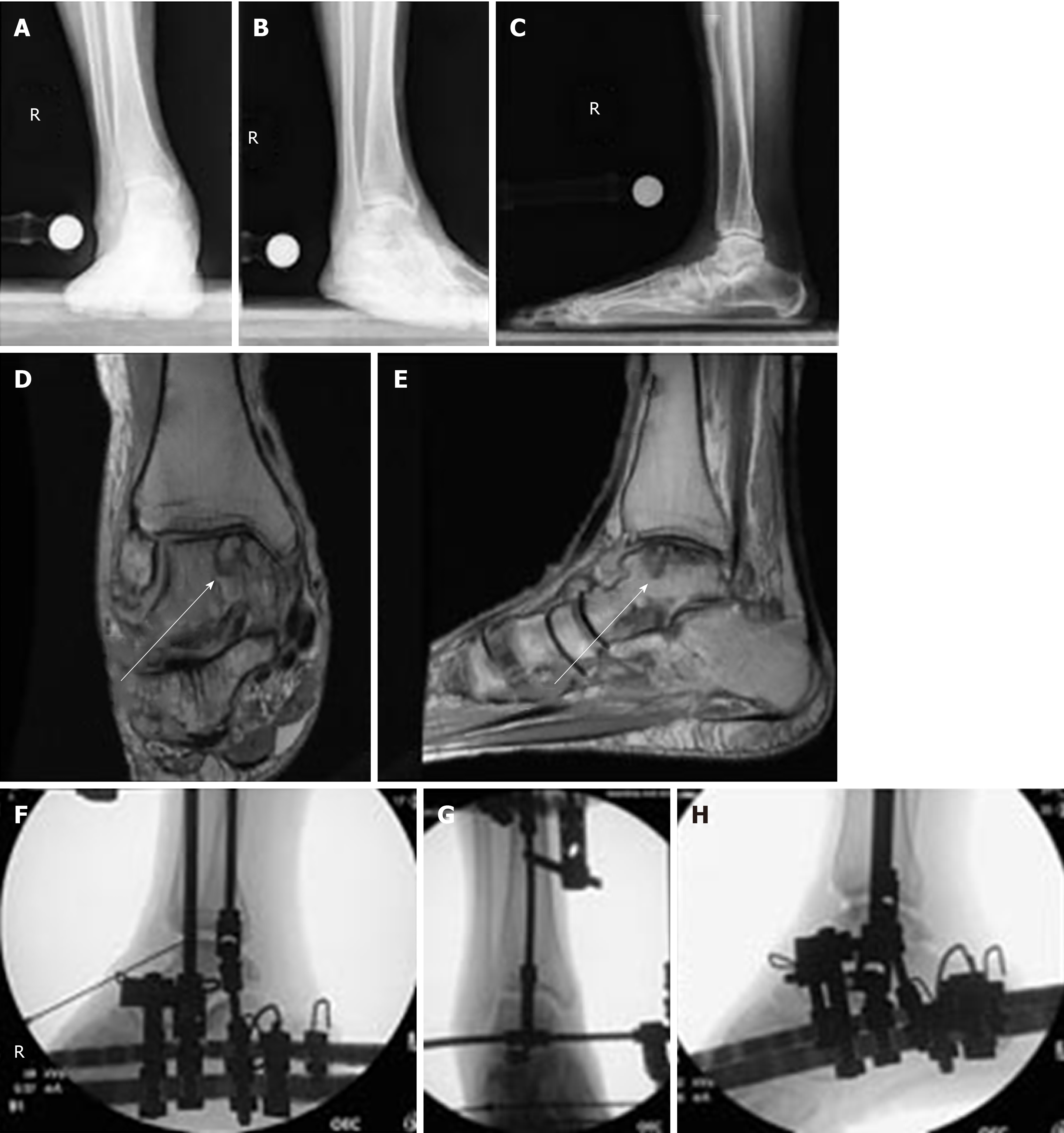
Figure 5 pre-operative standing X-rays and magnetic reconnaissance imaging, in addition to intra-operative fluoroscopy of the right ankle after anterior cheilectomy and distraction.
A: Anterior-Posterior (AP) X-rays demonstrates post-traumatic ankle osteoarthritis, joint space narrowing, anterior osteophyte, and flat foot deformity; B: Mortise view showing the same; C: Lateral view; D: Coronal view of the magnetic reconnaissance imaging shows medial osteochondritis dissecans lesion on the medial talar dome, as indicated by arrow; E: Sagittal view of the magnetic reconnaissance imaging showing medial osteochondritis dissecans lesion on the medial talar dome, as indicated by arrow; F: Anterior-Posterior intra-operative fluoroscopy of the right ankle after anterior cheilectomy, application of frame and distraction; G: Ankle in plantarflexion; H: Ankle in dorsiflexion with adjunctive injection of bone marrow aspirate concentrate into the ankle joint.
- Citation: Dabash S, Buksbaum JR, Fragomen A, Rozbruch SR. Distraction arthroplasty in osteoarthritis of the foot and ankle. World J Orthop 2020; 11(3): 145-157
- URL: https://www.wjgnet.com/2218-5836/full/v11/i3/145.htm
- DOI: https://dx.doi.org/10.5312/wjo.v11.i3.145


