Copyright
©The Author(s) 2020.
World J Orthop. Feb 18, 2020; 11(2): 129-136
Published online Feb 18, 2020. doi: 10.5312/wjo.v11.i2.129
Published online Feb 18, 2020. doi: 10.5312/wjo.v11.i2.129
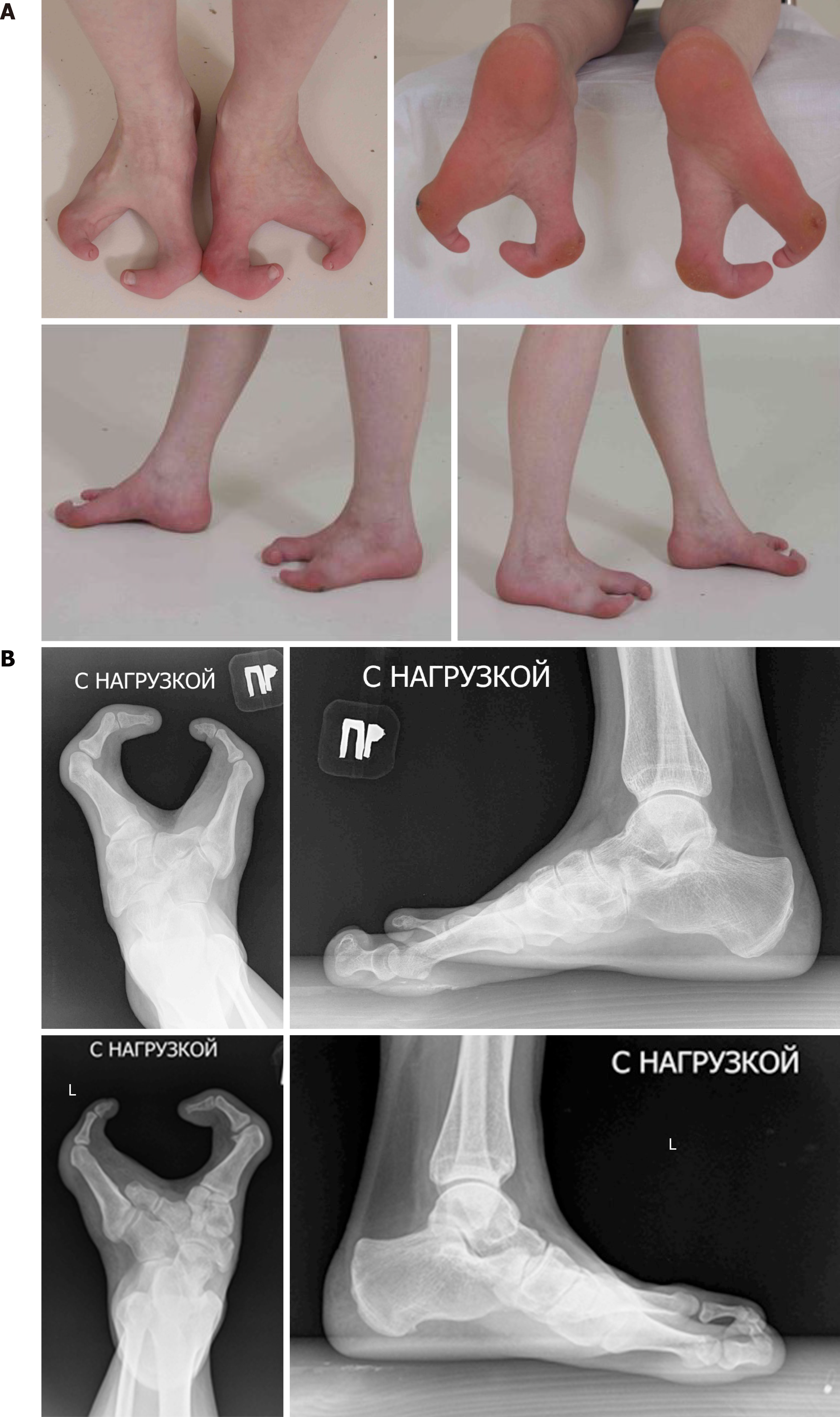
Figure 1 Photo and x-ray pictures of patient’s feet before treatment.
A: Cleft feet; B: X-rays of feet in anterior-posterior and lateral view (absence of central feet rays).
- Citation: Leonchuk SS, Neretin AS, Blanchard AJ. Cleft foot: A case report and review of literature. World J Orthop 2020; 11(2): 129-136
- URL: https://www.wjgnet.com/2218-5836/full/v11/i2/129.htm
- DOI: https://dx.doi.org/10.5312/wjo.v11.i2.129


