Copyright
©The Author(s) 2019.
World J Orthop. Apr 18, 2019; 10(4): 206-211
Published online Apr 18, 2019. doi: 10.5312/wjo.v10.i4.206
Published online Apr 18, 2019. doi: 10.5312/wjo.v10.i4.206
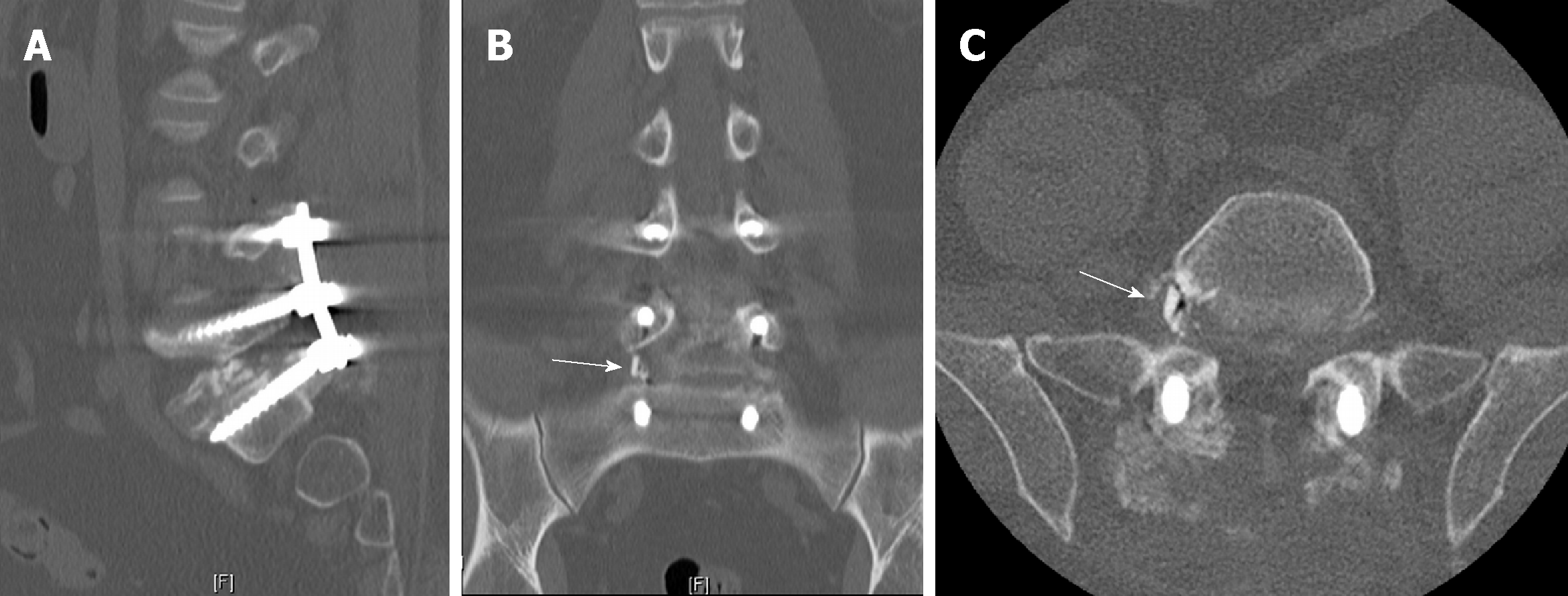
Figure 2 Postoperative computed tomography scan.
A: Sagittal view showed the main part of the allograft spacer in the disc space without clear view of broken fragment; B: Coronal view shows obvious fragment (arrow) in the right L5-S1 foramina; C: Axial view also shows obvious fragment (arrow) in the right L5-S1 foramina.
- Citation: Kyle A, Rowland A, Stirton J, Elgafy H. Fracture of allograft interbody spacer resulting in post-operative radiculopathy: A case report. World J Orthop 2019; 10(4): 206-211
- URL: https://www.wjgnet.com/2218-5836/full/v10/i4/206.htm
- DOI: https://dx.doi.org/10.5312/wjo.v10.i4.206


