
















©The Author(s) 2019.
World J Orthop. Jan 18, 2019; 10(1): 45-53
Published online Jan 18, 2019. doi: 10.5312/wjo.v10.i1.45
Published online Jan 18, 2019. doi: 10.5312/wjo.v10.i1.45
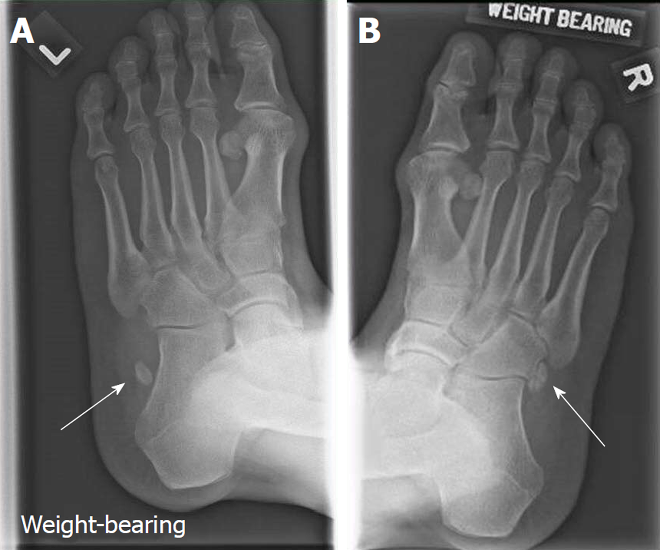
Figure 2 Comparing radiographs of bilateral oblique foot views.
A: Weight-bearing oblique view of left foot show a bony fragment lateral to the anterior left calcaneum; B: Shows the position of the undisplaced os peroneum on the right foot. The displaced os peroneum over the left foot is better appreciated when compared against the contralateral foot.
- Citation: Koh D, Liow L, Cheah J, Koo K. Peroneus longus tendon rupture: A case report. World J Orthop 2019; 10(1): 45-53
- URL: https://www.wjgnet.com/2218-5836/full/v10/i1/45.htm
- DOI: https://dx.doi.org/10.5312/wjo.v10.i1.45