Copyright
©The Author(s) 2018.
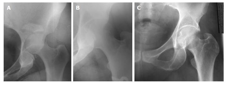
Figure 1 Plain radiographs of the pelvis both hip joints for a 4-year-old boy; the right hip joint was dislocated after minor trauma.
A: Notice the neck-shaft angle; B: A radiograph was taken one week after closed reduction shows the stable reduction; C: A radiograph was taken after three years of reduction shows no signs of avascular necrosis.
Figure 2 Plain radiographs of the pelvis both hip joints for a 27-year-old male, was admitted to hospital as a polytraumatized patient, the right hip dislocation was reduced two days after injury.
A: The right hip dislocated posteriorly; B: Stable reduction by the second month; C: A radiograph was taken 16 years after reduction shows avascular necrosis of the femoral head and degenerative changes of the right hip joint.
Figure 3 Plain radiographs of right hip joint for a 42-year-old female, was diagnosed two days after a motor car accident.
A: A posterior dislocation of the right hip with acetabular fractures; B: A radiograph was taken two weeks after closed reduction shows stable reduction; C: A radiograph was taken seven years after reduction shows no signs of avascular necrosis in the head of right femur.
- Citation: Massoud EIE. Neglected traumatic hip dislocation: Influence of the increased intracapsular pressure. World J Orthop 2018; 9(3): 35-40
- URL: https://www.wjgnet.com/2218-5836/full/v9/i3/35.htm
- DOI: https://dx.doi.org/10.5312/wjo.v9.i3.35


